
 By
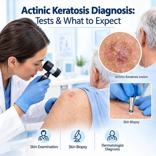
By Actinic keratosis diagnosis usually begins with a careful examination of the skin. A dermatologist looks closely at areas that have received significant sun exposure and checks for rough, scaly or discoloured patches. In many cases, a healthcare professional can identify actinic keratosis from its appearance and texture alone. However, a skin biopsy may be recommended when a lesion looks unusual, thick, persistent or potentially cancerous.
Knowing what happens during an actinic keratosis diagnosis can make the process less stressful. The examination is generally straightforward, and most people do not need extensive testing. Your dermatologist will assess your skin, ask about your history of sun exposure and previous skin problems, and decide whether further testing is necessary.
What Is Actinic Keratosis?
What Is Actinic Keratosis? Actinic keratosis is a common skin condition caused by long-term exposure to ultraviolet (UV) radiation. It usually develops on areas of skin that receive frequent sun exposure, such as the Actinic Keratosis on the Face, Actinic Keratosis on the Scalp, Actinic Keratosis on the Ears, neck, forearms and backs of the hands.
An actinic keratosis may appear as a small rough or scaly patch. The colour can vary from skin-coloured or pink to red, brown or grey. Some lesions are easier to feel than see because they have a sandpaper-like texture.
Actinic keratoses can occur as single lesions or as multiple patches. They are considered precancerous because some can develop into squamous cell carcinoma, a type of skin cancer. This does not mean that every actinic keratosis will become cancerous, but it makes proper assessment and monitoring important.
When Should You Seek an Actinic Keratosis Diagnosis?
You should consider having a persistent or suspicious skin lesion examined, particularly if it appears on an area that has received substantial sun exposure.
Common Early Signs of Actinic Keratosis that may warrant assessment include:
- A rough, scaly patch that does not disappear
- A spot that repeatedly becomes crusty
- A new area of persistent redness or scaling
- A lesion that feels rougher than the surrounding skin
- A patch that gradually becomes thicker
- A sore or crusted area that repeatedly returns
- A lesion that bleeds without an obvious injury
- A spot that becomes painful, tender or increasingly irritated
- A skin change that continues to grow or looks different from your other lesions
Some actinic keratoses are very small, while others may become several centimetres wide. Because early skin cancers can sometimes resemble actinic keratosis, an unusual lesion should not be diagnosed based only on photographs or appearance at home.
How Is Actinic Keratosis Diagnosed?
The main part of an actinic keratosis diagnosis is a physical examination of the skin. Your healthcare professional may also use a magnifying device called a dermatoscope to examine the lesion more closely.
A biopsy is not always necessary. Your dermatologist decides whether one is appropriate based on the lesion’s appearance, thickness, location and other clinical features.
The diagnosis generally involves three stages:
- Medical and skin history
- Detailed skin examination
- Additional testing when needed
Let’s look at each stage in more detail.
1. Your Medical and Skin History
Before examining your skin, your dermatologist may ask questions about your health and previous skin problems.
You may be asked about:
- How long you have noticed the lesion
- Whether it has changed in size or appearance
- Whether it itches, hurts or bleeds
- Your history of sun exposure
- Outdoor work or activities
- Use of tanning beds
- Previous sunburns
- Previous actinic keratoses
- Previous skin cancer
- Medications that may affect your immune system
- Family or personal history of skin conditions
Your dermatologist may also ask whether you have noticed other unusual areas elsewhere on your body.
This information helps place the lesion in context and can help determine whether additional testing is needed.
2. Skin Examination
A detailed skin examination is usually the most important part of the diagnostic process.
During the skin examination, your dermatologist will inspect the affected area and may examine surrounding skin for additional lesions. Actinic keratoses often occur in sun-exposed areas, and people can have more than one lesion without realising it.
Your dermatologist may look for:
- Scale
- Rough texture
- Changes in colour
- Redness
- Thickening
- Crusting
- Irregular borders
- Changes in size
- Tenderness or other symptoms
The doctor may also gently touch the lesion. Some actinic keratoses are easier to identify by texture than by appearance.
What Does a Dermatoscope Do?
A dermatoscope is a handheld device that provides magnification and specialised illumination. It allows a dermatologist to examine structures that may not be obvious to the naked eye.
Dermatoscopy can provide additional information when a lesion is difficult to classify from its surface appearance. It can also help a dermatologist distinguish some benign lesions from lesions that require further evaluation.
The examination is usually painless and does not involve cutting the skin.
3. Skin Biopsy
A skin biopsy may be recommended if the dermatologist cannot confidently distinguish actinic keratosis from another condition or if the lesion has features that raise concern for skin cancer.
A biopsy involves removing a small sample of skin and sending it to a laboratory. A pathologist examines the tissue under a microscope.
A biopsy may be particularly useful when a lesion is:
- Very thick
- Tender or painful
- Persistent despite treatment
- Bleeding or ulcerated
- Rapidly changing
- Unusually pigmented
- Suspicious for squamous cell carcinoma
A biopsy does not automatically mean that you have cancer. It simply provides tissue that can be examined more precisely.
What Happens During a Skin Biopsy?
If a skin biopsy is necessary, your healthcare professional will usually explain the procedure before starting.
The area is generally cleaned and numbed with a local anaesthetic. You may feel a brief sting from the anaesthetic injection, but the biopsy itself should not cause significant pain.
Depending on the lesion and the information needed, the dermatologist may use different biopsy techniques.
Shave Biopsy
A shave biopsy removes a thin layer of skin from the lesion. It may be appropriate for certain superficial lesions.
Punch Biopsy
A punch biopsy uses a small circular instrument to remove a deeper, cylindrical sample of skin. A stitch may sometimes be required.
Excisional Biopsy
An excisional biopsy removes the entire lesion or a larger portion of it. This approach may be considered when complete removal is appropriate or when a deeper examination is necessary.
The exact technique depends on the lesion and what your dermatologist needs to determine.
How Long Does an Actinic Keratosis Diagnosis Take?
The initial examination is usually relatively quick. If the dermatologist can confidently recognise the lesion as actinic keratosis, you may receive a diagnosis during the appointment.
A biopsy takes longer because the tissue must be processed and examined in a laboratory. The time required to receive the pathology result varies between healthcare providers and laboratories.
Your dermatologist should explain when you can expect the results and how they will be communicated.
Can Actinic Keratosis Be Diagnosed Without a Biopsy?
Yes. Many actinic keratoses have a characteristic appearance and texture, allowing an experienced dermatologist to make a clinical diagnosis without removing tissue.
However, appearance alone is not always enough.
Some skin cancers and other skin conditions can resemble actinic keratosis. A dermatologist may therefore recommend a biopsy when the lesion looks atypical or when there is uncertainty about the diagnosis.
The decision depends on the individual lesion rather than a requirement that every suspected actinic keratosis undergo a biopsy.
Actinic Keratosis vs Skin Cancer
One of the important reasons for an accurate actinic keratosis diagnosis is distinguishing it from skin cancer.
Actinic keratosis and squamous cell carcinoma can sometimes look similar. A lesion that becomes significantly thicker, painful, ulcerated or persistently crusted may require closer assessment.
You should tell your dermatologist if a suspected actinic keratosis has:
- Started growing quickly
- Become painful
- Developed significant thickness
- Repeatedly bled
- Developed an open sore
- Changed substantially in appearance
- Failed to improve after previous treatment
These signs do not prove that the lesion is cancerous. They indicate that further evaluation may be appropriate.
Can You Diagnose Actinic Keratosis at Home?
You can look for suspicious changes in your own skin, but self-diagnosis cannot reliably replace professional assessment.
A rough patch may be actinic keratosis, but similar changes can occur with eczema, psoriasis, seborrhoeic keratosis, warts, superficial skin infections and different types of skin cancer.
If you notice a persistent or changing lesion, arrange a professional assessment rather than relying solely on online photographs or descriptions.
Regular self-checks can still be useful. They help you notice new or changing areas that you can then discuss with a healthcare professional.
What Should You Tell Your Dermatologist?
Before your appointment, make a note of any skin changes you have noticed.
Useful information includes:
- When you first noticed the lesion
- Whether it has changed
- Any pain or tenderness
- Episodes of bleeding or crusting
- Previous treatments
- Previous actinic keratoses
- Previous skin cancer
- Your history of significant sun exposure
If you have several lesions, point them out during the examination. You may also want to ask whether you need a broader skin examination.
What Happens After the Diagnosis?
Once your dermatologist confirms actinic keratosis, they can discuss appropriate management. Treatment depends on factors such as the number of lesions, their location, thickness, Actinic Keratosis symptoms and your overall skin health.
Possible treatments may include:
- Cryotherapy
- Prescription topical medicines
- Photodynamic therapy
- Curettage or other minor procedures
- Other dermatologist-directed treatments
Your dermatologist may also recommend protecting your skin from further UV exposure.
Treatment is important because actinic keratoses can sometimes progress to squamous cell carcinoma. Your healthcare professional can help determine which lesions need treatment and whether you require ongoing skin checks.
Why Follow-Up Matters
Having one actinic keratosis can indicate significant cumulative sun damage. You may therefore develop additional lesions in the future.
Follow-up appointments can help your dermatologist identify new or changing lesions early. The frequency of follow-up depends on your individual Actinic Keratosis Risk Factors and previous skin findings.
Between appointments, continue checking your skin and report suspicious changes.
Protecting Your Skin After Diagnosis
Sun protection is an important part of reducing further UV damage.
Helpful measures include:
- Use a broad-spectrum sunscreen with an appropriate SPF
- Reapply sunscreen as directed, particularly during prolonged outdoor exposure
- Wear protective clothing
- Use a wide-brimmed hat when appropriate
- Seek shade during periods of strong sunlight
- Avoid tanning beds
- Take extra care during outdoor activities
Sun protection does not remove existing actinic keratoses, but it can help reduce additional UV exposure.
Frequently Asked Questions About Actinic Keratosis Diagnosis
Is actinic keratosis diagnosis painful?
The initial skin examination is generally painless. If you need a biopsy, local anaesthetic is normally used to numb the area. You may feel a brief sting when the anaesthetic is injected.
Does every actinic keratosis need a biopsy?
No. Many lesions can be diagnosed clinically during a skin examination. A biopsy is generally considered when the appearance is unusual or when the dermatologist needs to rule out another condition, including skin cancer.
Can a dermatologist diagnose actinic keratosis immediately?
Often, yes. An experienced dermatologist may recognise a typical actinic keratosis during the examination. If the lesion is uncertain or suspicious, further testing may be recommended.
What does actinic keratosis feel like?
It often feels rough, dry or scaly. Some lesions have a texture similar to fine sandpaper. A lesion can sometimes be easier to feel than see.
Is actinic keratosis cancer?
Actinic keratosis is generally considered a precancerous skin lesion rather than skin cancer. Some lesions can progress to squamous cell carcinoma, so professional assessment and appropriate management are important.
What happens if a biopsy shows skin cancer?
If pathology identifies skin cancer, your dermatologist will discuss the diagnosis and treatment options with you. The next steps depend on the specific type, location, size and extent of the cancer.
Final Thoughts
An actinic keratosis diagnosis usually starts with a straightforward skin examination. Your dermatologist assesses the lesion’s appearance and texture and may use dermatoscopy to examine it in greater detail. Most typical lesions can be diagnosed without extensive testing.
When a lesion looks unusual, thick, persistent or suspicious, a skin biopsy can provide a more definite answer. The biopsy allows a pathologist to examine the tissue under a microscope and determine whether the lesion is actinic keratosis or another skin condition.
A timely dermatologist diagnosis can help identify actinic keratoses and distinguish suspicious lesions from skin cancer. If you notice a rough, scaly or changing patch that does not go away, professional assessment is the safest way to determine what it represents.
Medical Disclaimer
This article is provided for general educational purposes and should not replace professional medical advice, diagnosis or treatment. If you have a new, persistent, changing, painful or bleeding skin lesion, consult a qualified healthcare professional or dermatologist for an appropriate evaluation.