
 By
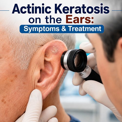
By Actinic Keratosis on the Ears is a common sun-related skin condition that develops when years of ultraviolet (UV) exposure damage skin cells. The ears are particularly vulnerable because they are often exposed to sunlight but may be overlooked when people apply sunscreen.
Actinic keratosis (AK), also called solar keratosis, usually appears as a rough, dry or scaly patch. It may be skin-coloured, pink, red, brown, grey or whitish. Some lesions cause itching, tenderness, burning or pain, while others produce no noticeable symptoms.
Although actinic keratosis is not itself invasive skin cancer, it is considered a precancerous skin change because some lesions can develop into squamous cell carcinoma (SCC). For this reason, persistent or changing ear skin lesions should be assessed by a doctor or dermatologist. The Actinic Keratosis symptoms, causes, risk factors, diagnosis, treatment options and prevention of Actinic Keratosis on the Ears.
What is Actinic Keratosis on the Ears?
What is Actinic Keratosis on the Ears? Actinic keratosis occurs when repeated UV exposure causes abnormal changes in skin cells. These changes typically develop gradually over years rather than appearing after a single day in the sun.
The ears are common locations for actinic keratoses because the outer ear, particularly the helix and other exposed surfaces, receives considerable sunlight. People who spend substantial time outdoors, especially without adequate sun protection, may develop multiple lesions.
An actinic keratosis may initially look like a small area of rough or dry skin. In some cases, you may notice the roughness by touch before the lesion becomes obvious visually.
AKs can occur as individual spots or as several patches across chronically sun-damaged skin. They may also recur after treatment because the surrounding skin may have accumulated significant UV damage.
What Causes Actinic Keratosis on the Ears?
The primary cause is long-term exposure to ultraviolet radiation, particularly UVB. UV radiation can damage the DNA within skin cells. Over time, repeated damage can lead to abnormal cell growth and the development of actinic keratoses.
Common sources of UV exposure include:
- Direct sunlight
- Repeated sunburn
- Long hours spent outdoors
- Outdoor occupations
- Recreational activities in the sun
- Indoor tanning beds and sunlamps
- Inadequate use of sunscreen or protective clothing
The risk increases with cumulative exposure, meaning that years of sun exposure can contribute to AK even if a person does not remember having frequent sunburns.
The ears can be especially vulnerable because they are easy to overlook when applying sunscreen. Hats can provide additional protection, but many hats do not completely cover the ears.
Symptoms of Actinic Keratosis on the Ears
The appearance of Actinic Keratosis on the Ears can vary considerably. Some lesions are subtle, while others become thick, crusty or clearly visible.
Common Actinic Keratosis symptoms and signs include:
1. Rough or Sandpaper-Like Skin
One of the earliest Signs of Actinic Keratosis may be a rough patch that feels similar to sandpaper. You might notice the texture when running your finger over the ear even when the lesion is difficult to see.
2. Dry, Scaly Patches
Actinic keratoses often develop a dry or scaly surface. The scale may be white, yellowish, grey or skin-coloured.
3. Pink or Red Areas
Some AKs appear as pink or red patches, particularly on lighter skin. The surrounding skin may also show signs of chronic sun damage.
4. Brown, Tan or Skin-Coloured Lesions
Actinic keratoses do not always appear red. They may also be tan, brown, grey or close to the person’s natural skin tone.
5. Itching or Irritation
An AK may itch or feel irritated. However, the absence of symptoms does not necessarily mean that a lesion is harmless.
6. Burning, Stinging or Tenderness
Some people experience burning, stinging or tenderness, particularly when the lesion is touched or exposed to friction.
7. Thickening or Crusting
An actinic keratosis can become thicker over time. A heavily thickened lesion may develop a hard or crusted surface.
8. Bleeding
Occasional bleeding can occur, particularly if a rough lesion is scratched or irritated. However, repeated bleeding or spontaneous bleeding should be assessed by a healthcare professional.
The NHS recommends medical assessment when an actinic keratosis becomes larger, changes colour, becomes tender, bleeds or develops into a lump.
What Does Actinic Keratosis on the Ear Look Like?
The appearance can range from a barely noticeable rough spot to a clearly visible scaly lesion.
You may notice:
- A small rough patch
- A dry, flaky area
- A pink or red spot
- A brown or tan patch
- A thickened scaly growth
- A crusty area
- A horn-like projection in more pronounced lesions
Because different skin conditions can produce similar-looking patches, you should not rely on appearance alone to diagnose an ear lesion.
Other conditions can cause ear skin lesions, including eczema, psoriasis, seborrhoeic keratosis, basal cell carcinoma and squamous cell carcinoma. A dermatologist can distinguish these conditions through examination and, when necessary, dermoscopy or biopsy.
Who Is at Higher Risk?
Anyone can develop actinic keratosis after sufficient UV exposure, but certain factors increase the likelihood.
Risk factors include:
- Older age
- A history of significant sun exposure
- Frequent sunburn
- Fair or sun-sensitive skin
- Working outdoors
- Spending considerable time outdoors for recreation
- Previous actinic keratoses
- A weakened immune system
- Previous skin cancer
- Regular exposure to tanning beds or sunlamps
People who live in areas with strong sunlight or spend many years outdoors may accumulate substantial UV damage.
Importantly, having an actinic keratosis indicates that the skin has experienced significant UV damage. It also means that additional actinic keratoses and other types of skin cancer may develop in the future.
Is Actinic Keratosis on the Ears Cancer?
No. Actinic keratosis is not invasive skin cancer. However, it is considered a precancerous lesion because some AKs can progress to squamous cell carcinoma.
Squamous cell carcinoma is a type of skin cancer that can occur on chronically sun-damaged areas, including the ears. The risk associated with an individual AK is generally low, but the presence of AK indicates significant cumulative UV damage.
This is why doctors may recommend treating actinic keratoses rather than simply ignoring them.
Signs That May Suggest Squamous Cell Carcinoma
A lesion should receive prompt medical assessment if it:
- Grows quickly
- Becomes increasingly thick
- Develops significant tenderness or pain
- Bleeds repeatedly
- Forms a persistent ulcer
- Develops a firm lump
- Becomes increasingly crusted
- Changes noticeably in appearance
- Fails to heal
A rapidly growing, thick, painful or ulcerated lesion may require biopsy to rule out SCC.
How Is Actinic Keratosis on the Ears Diagnosed?
A doctor or dermatologist usually begins with a visual examination and medical history.
They may ask about:
- How long the lesion has been present
- Whether it has changed
- Previous sun exposure
- History of sunburn
- Previous skin cancers
- Symptoms such as pain, bleeding or itching
- Previous treatment for actinic keratosis
A dermatologist may use a dermatoscope, a specialised magnifying device that helps examine skin structures more closely.
In some cases, a biopsy may be recommended. This is particularly important when the lesion looks unusual, becomes thick or painful, bleeds, continues to grow or does not respond appropriately to treatment.
Actinic Keratosis Treatment Options
The appropriate Actinic Keratosis treatment depends on the number, size, thickness and location of lesions, as well as the patient’s medical history and the dermatologist’s assessment.
Cryotherapy
Cryotherapy uses liquid nitrogen to freeze and destroy the abnormal skin cells.
It is commonly used when there are one or a few individual lesions. After treatment, the area may become red, swollen or blistered before healing.
Healing time varies according to the treatment intensity and location.
Prescription Creams and Gels
Topical medicines may be prescribed when several AKs are present or when there is a broader area of sun-damaged skin.
Depending on the clinical situation, treatments may include:
- 5-fluorouracil
- Imiquimod
- Diclofenac
- Tirbanibulin
These medicines work in different ways to eliminate or treat abnormal cells. Some cause noticeable redness, scaling, inflammation or irritation during treatment, so they should be used exactly as prescribed.
Photodynamic Therapy
Photodynamic therapy (PDT) combines a photosensitising medication with a specific light source.
The medication is applied to the affected area and activated with light, helping destroy abnormal cells. PDT can be particularly useful when there are multiple lesions or areas of sun-damaged skin.
Curettage and Electrosurgery
For thicker or more pronounced lesions, a dermatologist may scrape away abnormal tissue using curettage and may use electrosurgery to destroy remaining abnormal cells.
This approach can also provide tissue for laboratory examination when cancer needs to be excluded.
Surgical Removal
Some suspicious or particularly thick lesions may require surgical excision or another procedure.
If squamous cell carcinoma is suspected, surgical management may be more appropriate than routine AK treatment. The removed tissue can then be examined under a microscope.
The choice of procedure depends on the lesion’s characteristics and the dermatologist’s assessment.
Can Actinic Keratosis on the Ears Go Away on Its Own?
Yes, some actinic keratoses can spontaneously regress. However, this does not mean that every rough patch should simply be ignored.
The underlying sun damage remains, and new lesions can develop. Some AKs can also persist or progress.
For this reason, monitoring your skin and discussing persistent or changing lesions with a healthcare professional is important.
How to Prevent Sun Damage on Ears
Preventing additional sun damage on ears is an important part of managing actinic keratosis.
Apply Broad-Spectrum Sunscreen
Use a broad-spectrum sunscreen with an SPF of at least 30 on exposed ears. Apply it generously and reapply as directed, particularly during prolonged outdoor activities.
Do not forget the tops, sides and other exposed surfaces of the ears.
Wear a Wide-Brimmed Hat
A wide-brimmed hat can provide additional protection for the ears, Actinic Keratosis on the Lips, Actinic Keratosis on the Scalp, Actinic Keratosis on the Face and neck. A cap may leave the ears exposed, so consider the type of hat you wear during prolonged sun exposure.
Seek Shade
Reducing direct exposure to intense sunlight can help minimise cumulative UV damage.
Avoid Tanning Beds
Indoor tanning exposes the skin to UV radiation and can contribute to skin damage and skin cancer risk.
Check Your Ears Regularly
Because the ears are easy to overlook, include them when performing a regular skin check.
Use a mirror or ask someone you trust to look at areas that are difficult for you to see.
Look for:
- New rough patches
- Persistent scaling
- Bleeding spots
- Crusting
- New lumps
- Changes in existing lesions
- Areas that repeatedly become irritated
When Should You See a Dermatologist?
You should arrange a medical assessment if you notice a new or persistent lesion on your ear, particularly if it changes over time.
Seek professional advice if an area:
- Becomes thicker
- Grows
- Changes colour
- Bleeds
- Becomes painful or tender
- Develops a lump
- Becomes ulcerated
- Does not heal
- Returns after treatment
This is particularly important because the ears are a recognised site for sun-related skin cancers, including squamous cell carcinoma.
Do not attempt to remove a suspected actinic keratosis at home by cutting, scraping, burning or using unprescribed chemicals. A dermatologist can determine the diagnosis and recommend an appropriate treatment.
Frequently Asked Questions
Can actinic keratosis develop on the ears?
Yes. The ears are common sites for actinic keratoses because they are frequently exposed to UV radiation.
Is Actinic Keratosis on the Ears dangerous?
Actinic keratosis is not invasive cancer, but it is a precancerous skin change. Some lesions can develop into squamous cell carcinoma, so persistent or changing lesions should be assessed.
Does actinic keratosis hurt?
It may be completely asymptomatic, but some lesions can itch, burn, sting or become tender and painful.
What is the best Actinic Keratosis treatment?
There is no single treatment that is best for everyone. Cryotherapy, prescription topical treatments, photodynamic therapy and procedures such as curettage may all be appropriate depending on the lesion and the patient’s circumstances.
Can sunscreen prevent actinic keratosis?
Consistent sun protection can reduce UV-related skin damage and help reduce the development and symptoms of actinic keratoses. Sunscreen should be applied to exposed areas, including the ears.
Final Thoughts
Actinic Keratosis on the Ears develops as a result of cumulative UV damage and commonly appears as a rough, dry, scaly or discoloured patch. Although it is not invasive skin cancer, it can sometimes progress to squamous cell carcinoma.
The ears deserve particular attention because they are frequently exposed to sunlight and are often missed during routine sunscreen application. Early assessment of suspicious ear skin lesions can help distinguish actinic keratosis from other skin conditions and identify lesions that may require more urgent investigation.
Treatment may involve cryotherapy, prescription creams or gels, photodynamic therapy, curettage or surgical removal, depending on the lesion. Regular skin checks and consistent protection from UV radiation are also important for reducing further sun damage on ears.
If you notice a rough, thickened, painful, bleeding or changing spot on your ear, arrange an examination with a qualified healthcare professional rather than attempting to diagnose or treat it yourself.
Medical Disclaimer
This article is for general educational purposes only and does not provide a diagnosis or personalised medical advice. Actinic keratosis can resemble other skin conditions, including skin cancer. If you have a new, changing, bleeding, painful or persistent lesion on your ear, consult a qualified doctor or dermatologist for appropriate evaluation and treatment.