 By
By Receiving a new skin growth can be concerning, especially when it changes in appearance or resembles more serious conditions such as skin cancer. Fortunately, many skin growths are harmless, and one of the most common examples is seborrheic keratosis. Although these growths are benign, proper seborrheic keratosis diagnosis is important to ensure that the lesion is not another skin condition requiring treatment.
Seborrheic keratosis affects millions of adults worldwide, particularly those over the age of 40. These growths often appear as brown, black, or tan spots with a waxy or wart-like texture. While experienced dermatologists can frequently identify them through visual examination alone, additional diagnostic tools may sometimes be necessary. The complete process of seborrheic keratosis diagnosis, including skin examinations, dermoscopy, biopsy procedures, differential diagnosis, and when medical evaluation is recommended.
What is Seborrheic Keratosis?
Seborrheic keratosis is a noncancerous skin growth that develops from the outer layer of the skin. These lesions can appear almost anywhere on the body except the palms and soles.
Common characteristics include:
- Brown, black, yellow, or tan coloration
- Waxy or rough texture
- Slightly raised appearance
- Round or oval shape
- “Stuck-on” look
- Slow growth over time
They commonly appear on:
- Seborrheic Keratosis on Face
- Chest
- Back
- Neck
- Shoulders
- Scalp
Although harmless, they can resemble other skin conditions, making accurate diagnosis essential.
Why Is Seborrheic Keratosis Diagnosis Important?
Many skin cancers and precancerous lesions can look similar to seborrheic keratosis during early stages.
A proper seborrheic keratosis diagnosis helps:
- Confirm the growth is benign
- Rule out melanoma
- Exclude basal cell carcinoma
- Distinguish from actinic keratosis
- Determine whether Seborrheic Keratosis Treatment is necessary
- Reduce unnecessary anxiety
While seborrheic keratosis itself is not dangerous, any unusual skin growth deserves professional evaluation if there is uncertainty about its nature.
The First Step: Clinical Skin Examination
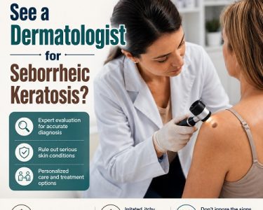
A visual skin examination is usually the first stage of seborrheic keratosis diagnosis.
Medical History Review
A dermatologist may ask questions such as:
- When did the growth first appear?
- Has it changed recently?
- Is it itchy or painful?
- Does it bleed?
- Have similar lesions appeared elsewhere?
- Is there a personal or family history of skin cancer?
These details provide important clues regarding the lesion’s behavior.
Visual Assessment
During examination, doctors evaluate:
Size
Seborrheic keratoses range from a few millimeters to several centimeters.
Color
They may appear:
- Light tan
- Brown
- Dark brown
- Black
Texture
Most lesions have:
- Waxy surfaces
- Rough texture
- Wart-like appearance
Border
Seborrheic keratoses usually have well-defined borders.
Distribution
Multiple lesions appearing over time strongly suggest seborrheic keratosis.
Typical Features Doctors Look For
Experienced dermatologists often recognize seborrheic keratosis immediately because of its distinctive appearance.
Classic signs include:
- Raised plaque-like lesion
- Stuck-on appearance
- Greasy or waxy surface
- Sharp edges
- Uniform color pattern
- Slow growth
When these features are present, diagnosis can often be made without additional testing.
What Is Dermoscopy?
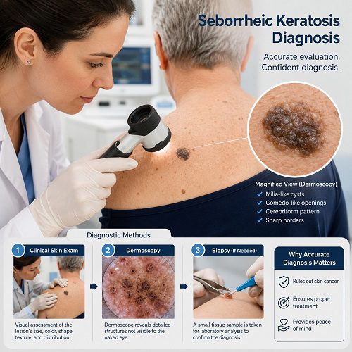
Dermoscopy is one of the most valuable tools in modern dermatology.
A dermoscope is a handheld device equipped with:
- Magnification
- Specialized lighting
- Polarized lenses
This instrument allows dermatologists to see structures beneath the skin surface that are invisible to the naked eye.
Dermoscopy significantly improves the accuracy of seborrheic keratosis diagnosis.
How Dermoscopy Works
The dermatologist places the dermoscope directly over the lesion.
The device magnifies the skin and reveals:
- Pigment patterns
- Surface structures
- Blood vessels
- Keratin features
These details help distinguish benign lesions from potentially cancerous ones.
Dermoscopic Features of Seborrheic Keratosis
Several dermoscopic findings strongly suggest seborrheic keratosis.
Milia-Like Cysts
These appear as:
- Small white or yellow dots
- Tiny keratin-filled cysts
They are among the most recognizable dermoscopic signs.
Comedo-Like Openings
These resemble:
- Small black pores
- Tiny surface pits
They result from keratin accumulation.
Fissures and Ridges
Dermoscopy often reveals:
- Brain-like grooves
- Surface ridges
- Cerebriform patterns
These features are highly characteristic.
Sharp Borders
Seborrheic keratoses usually demonstrate clearly defined margins under dermoscopy.
Moth-Eaten Borders
Some lesions show irregular but benign-appearing edges known as moth-eaten borders.
Advantages of Dermoscopy
Dermoscopy offers several benefits:
- Noninvasive
- Quick procedure
- Painless
- No recovery required
- Improved diagnostic accuracy
- Better melanoma detection
Many dermatologists routinely use dermoscopy during skin evaluations.
When Dermoscopy Is Not Enough
Occasionally, a lesion has unusual features.
Examples include:
- Rapid growth
- Irregular pigmentation
- Bleeding
- Ulceration
- Inflammation
- Uncertain appearance
In these cases, a biopsy may be recommended.
What Is a Skin Biopsy?
A skin biopsy involves removing a small sample of tissue for microscopic examination.
Biopsy is considered the gold standard when diagnosis is uncertain.
Although most seborrheic keratoses do not require biopsy, it becomes necessary when skin cancer cannot be confidently ruled out.
Reasons for Biopsy
Doctors may recommend biopsy if:
- The lesion is unusually dark
- Color varies significantly
- Borders are irregular
- The lesion changes rapidly
- Bleeding occurs without injury
- Seborrheic Keratoses Symptoms are unusual
- Dermoscopy findings are inconclusive
Biopsy provides definitive confirmation.
Types of Skin Biopsies
Several biopsy methods may be used.
Shave Biopsy
The most common approach.
The doctor:
- Numbs the skin
- Removes the top portion of the lesion
- Sends tissue for laboratory analysis
Advantages include:
- Quick procedure
- Minimal scarring
- Fast healing
Punch Biopsy
A circular tool removes a deeper tissue sample.
This method may be used when deeper evaluation is required.
Excisional Biopsy
The entire lesion is removed.
This is typically reserved for lesions strongly suspected of being cancerous.
What Happens During a Biopsy?
The procedure usually takes less than 30 minutes.
Steps include:
- Cleaning the area
- Local anesthesia injection
- Tissue removal
- Bleeding control
- Dressing application
- Laboratory analysis
Most patients experience little discomfort.
Pathology Examination
After biopsy, a dermatopathologist examines the tissue under a microscope.
This analysis confirms whether the lesion is:
- Seborrheic keratosis
- Melanoma
- Basal cell carcinoma
- Squamous cell carcinoma
- Actinic keratosis
- Another skin condition
Pathology provides the final diagnosis.
Microscopic Features of Seborrheic Keratosis
Under the microscope, seborrheic keratosis typically shows:
- Thickened epidermis
- Excess keratin production
- Horn cysts
- Benign cellular architecture
- No cancerous invasion
These findings confirm the diagnosis.
Conditions That Can Mimic Seborrheic Keratosis
Several skin disorders can resemble seborrheic keratosis.
Melanoma
Melanoma is the most important condition to rule out.
Warning signs include:
- Multiple colors
- Asymmetry
- Irregular borders
- Rapid changes
Some melanomas closely resemble seborrheic keratoses.
Actinic Keratosis
Actinic keratosis is a precancerous lesion caused by sun damage.
Features include:
- Rough patches
- Scaling
- Redness
These lesions require monitoring and treatment.
Basal Cell Carcinoma
Basal cell carcinoma may appear:
- Shiny
- Pink
- Pigmented
Dermoscopy helps differentiate it from seborrheic keratosis.
Squamous Cell Carcinoma
This cancer can present as:
- Thickened plaques
- Crusted lesions
- Non-healing sores
Biopsy may be necessary.
Common Warts
Warts share some similarities but often have:
- Different vascular patterns
- Viral origins
- Distinct dermoscopic findings
The Role of Artificial Intelligence in Diagnosis
Modern dermatology increasingly incorporates artificial intelligence (AI).
AI-assisted imaging systems can:
- Analyze lesion photographs
- Detect suspicious features
- Support dermatologist evaluations
However, AI does not replace professional examination and biopsy when needed.
Diagnosing Seborrheic Keratosis in Older Adults
Seborrheic keratoses become increasingly common with age.
In older adults, dermatologists may see:
- Numerous lesions
- Variable pigmentation
- Large growths
Because skin cancer risk also rises with age, thorough evaluation remains important.
Sudden Appearance of Multiple Lesions
A sudden eruption of many seborrheic keratoses may occasionally indicate the Leser-Trélat sign.
Although rare, this phenomenon can sometimes be associated with internal malignancies.
Doctors may investigate further if numerous lesions appear rapidly.
Can You Diagnose Seborrheic Keratosis at Home?
Self-assessment may provide clues, but definitive diagnosis should come from a healthcare professional.
Many skin cancers resemble benign lesions.
Avoid relying solely on:
- Internet images
- Online Seborrheic Keratosis symptoms checkers
- Self-diagnosis apps
Professional evaluation remains the safest approach.
When Should You See a Dermatologist?
Seek medical evaluation if a lesion:
- Changes rapidly
- Bleeds
- Becomes painful
- Develops irregular borders
- Shows multiple colors
- Appears suddenly
- Causes concern
Early assessment helps rule out serious conditions.
What Happens After Diagnosis?
Once seborrheic keratosis diagnosis is confirmed, treatment may not be necessary.
Many patients simply monitor the lesion.
Removal may be considered if the growth:
- Seborrheic Keratosis Causes irritation
- Snags on clothing
- Bleeds from friction
- Creates cosmetic concerns
Treatment options include:
- Cryotherapy
- Curettage
- Electrocautery
- Laser therapy
- Surgical removal
Prognosis After Seborrheic Keratosis Diagnosis
The outlook is excellent.
Seborrheic keratosis:
- Is benign
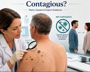
- Does not spread
- Rarely causes complications
- Has no cancerous potential in most cases
Regular skin checks can help monitor for new lesions and identify unrelated skin cancers early.
Preventing Seborrheic Keratosis Be Misdiagnosed
Preventing Seborrheic Keratosis Be Misdiagnosed: To ensure accurate diagnosis:
- Schedule regular skin examinations
- Monitor new skin growths
- Photograph changing lesions
- Use sun protection
- Consult a dermatologist when uncertain
Early evaluation prevents unnecessary worry and promotes timely treatment if another condition is discovered.
Frequently Asked Questions (FAQ) About Seborrheic Keratosis Diagnosis
1. What is seborrheic keratosis diagnosis?
Seborrheic keratosis diagnosis is the process healthcare professionals use to identify and confirm that a skin growth is a benign seborrheic keratosis rather than another skin condition, such as melanoma or other forms of skin cancer.
2. How do dermatologists diagnose seborrheic keratosis?
Dermatologists usually diagnose seborrheic keratosis through a visual skin examination. They assess the lesion’s color, texture, size, shape, and location. In many cases, the characteristic “stuck-on” appearance is enough for diagnosis.
3. What is dermoscopy, and why is it used?
Dermoscopy is a non-invasive diagnostic technique that uses a handheld device called a dermoscope to magnify and illuminate the skin. It helps dermatologists see structures beneath the skin surface and improves the accuracy of seborrheic keratosis diagnosis.
4. Is a biopsy always needed for seborrheic keratosis diagnosis?
No. Most cases can be diagnosed through a skin examination and dermoscopy. A biopsy is usually only recommended if the lesion has unusual features or if skin cancer cannot be confidently ruled out.
5. What happens during a skin biopsy?
During a biopsy, the area is numbed with a local anesthetic, and a small sample or the entire lesion is removed. The tissue is then sent to a laboratory for microscopic examination to confirm the diagnosis.
6. Can seborrheic keratosis be mistaken for skin cancer?
Yes. Seborrheic keratosis can sometimes resemble melanoma, basal cell carcinoma, or other skin cancers. This is why professional evaluation is important for any suspicious or changing skin lesion.
7. What does seborrheic keratosis look like?
Seborrheic keratosis usually appears as a brown, black, or tan growth with a waxy, rough, or wart-like surface. It often looks as though it is sitting on top of the skin.
8. Is dermoscopy painful?
No. Dermoscopy is completely painless and non-invasive. The dermatologist simply places the dermoscope against the skin to examine the lesion in greater detail.
9. How long does it take to receive biopsy results?
Biopsy results are typically available within a few days to two weeks, depending on the laboratory and healthcare provider.
10. When should I see a dermatologist for a skin lesion?
You should see a dermatologist if a skin growth changes in size, shape, or color, becomes painful, bleeds, itches excessively, or develops irregular borders. Any new or unusual lesion should be professionally evaluated.
11. Can I diagnose seborrheic keratosis myself?
While online images and resources may help you recognize common features, self-diagnosis is not recommended. Many skin cancers can resemble seborrheic keratosis, making a professional examination essential.
12. What happens after seborrheic keratosis diagnosis is confirmed?
Once seborrheic keratosis diagnosis is confirmed, treatment is often unnecessary because the growth is benign. However, it can be removed if it becomes irritated, bothersome, or cosmetically undesirable.
13. Does seborrheic keratosis turn into skin cancer?
Seborrheic keratosis is generally considered a benign condition and does not usually become cancerous. However, any suspicious changes should still be evaluated by a dermatologist.
14. Can multiple seborrheic keratoses appear at the same time?
Yes. Many people develop multiple seborrheic keratoses over time, especially as they age. In rare cases, a sudden eruption of numerous lesions may require further medical investigation.
15. What is the most accurate method for seborrheic keratosis diagnosis?
A skin biopsy is considered the most definitive diagnostic method because it allows microscopic examination of the tissue. However, in most cases, a dermatologist can accurately diagnose seborrheic keratosis through clinical examination and dermoscopy alone.
Conclusion
A proper seborrheic keratosis diagnosis involves a combination of clinical examination, dermoscopy, and occasionally biopsy. Most cases can be identified through their characteristic waxy, raised, and “stuck-on” appearance. Dermoscopy provides additional detail that improves diagnostic accuracy, while biopsy serves as the definitive test when uncertainty remains.
Although seborrheic keratosis is harmless, many skin cancers can mimic its appearance. For this reason, any changing, bleeding, or unusual skin lesion should be evaluated by a dermatologist. Early and accurate diagnosis offers peace of mind and ensures that potentially serious skin conditions are not overlooked.