 By
By Can Seborrheic Keratosis Be Misdiagnosed?
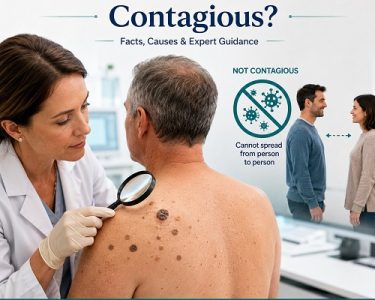
Seborrheic keratosis is one of the most common noncancerous skin growths seen in adults, particularly after the age of 40. These growths often appear as waxy, rough, or slightly raised patches that seem to be “stuck on” the surface of the skin. Although seborrheic keratoses are harmless, they can closely resemble several other skin conditions, including some forms of skin cancer.
Because of these similarities, seborrheic keratosis misdiagnosis can occur. A harmless growth may be mistaken for a dangerous lesion, or more concerningly, a skin cancer may initially be mistaken for seborrheic keratosis. This is why it is important not to rely solely on appearance when evaluating new or changing skin growths.
What Is Seborrheic Keratosis?
What Is Seborrheic Keratosis, Seborrheic keratosis is a benign skin growth that develops from the outer layer of the skin. It commonly appears on the:
- Chest
- Back
- Face
- Neck
- Shoulders
- Abdomen
These growths can vary greatly in appearance. Some are flat, while others are raised. Their color ranges from light tan to dark brown or nearly black.
Common characteristics include:
- Waxy appearance
- Rough or wart-like texture
- Round or oval shape
- Clearly defined borders
- Slow growth over time
- Usually painless
Although they are harmless, they may become irritated by clothing, shaving, or scratching.
Can Seborrheic Keratosis Be Misdiagnosed?
Yes. Seborrheic keratosis misdiagnosis is possible because many skin conditions share similar visual characteristics.
Even experienced clinicians sometimes require specialized tools or a biopsy to confirm the diagnosis. While many seborrheic keratoses have classic features, atypical lesions can closely resemble malignant tumors.
Misdiagnosis can occur in two ways:
- A benign seborrheic keratosis is mistaken for skin cancer, leading to unnecessary worry or Treatment for Seborrheic keratosis.
- A skin cancer is mistaken for seborrheic keratosis, delaying appropriate diagnosis and management.
The second scenario is particularly important because early detection significantly improves outcomes for many skin cancers.
Why Is Seborrheic Keratosis Difficult to Diagnose?
Several factors contribute to diagnostic confusion.
Wide Variation in Appearance
Seborrheic keratoses do not all look alike. Some are:
- Flat
- Thick
- Crusty
- Darkly pigmented
- Light-colored
- Inflamed
This variability increases the likelihood of confusion.
Skin Tone Differences
The appearance of seborrheic keratosis may differ depending on a person’s skin tone. Pigmented lesions can be especially difficult to distinguish from melanoma in individuals with darker skin.
Irritation
Repeated rubbing, scratching, or trauma may Causes of Seborrheic Keratosis to become:
- Red
- Bleeding
- Swollen
- Crusted
These changes can mimic skin cancer.
Multiple Lesions
People often develop numerous seborrheic keratoses with age. While this pattern is typical, new or unusual lesions among existing growths deserve careful evaluation.
Conditions That Can Be Mistaken for Seborrheic Keratosis
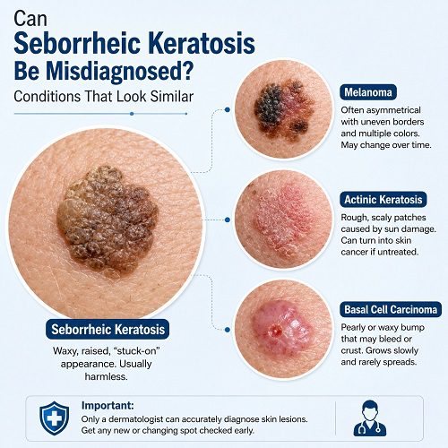
Several skin conditions resemble seborrheic keratosis.
1. Melanoma
Melanoma is the most serious condition that may resemble seborrheic keratosis.
Both may appear:
- Dark brown
- Black
- Raised
- Irregular
However, melanoma often demonstrates warning signs including:
- Asymmetry
- Irregular borders
- Multiple colors
- Diameter larger than 6 mm
- Evolution over time
Some melanomas imitate seborrheic keratoses so closely that even dermatologists may perform a biopsy to confirm the diagnosis.
2. Basal Cell Carcinoma
Basal cell carcinoma is the most common skin cancer.
Certain pigmented forms may resemble seborrheic keratosis because they:
- Grow slowly
- Develop raised edges
- Become crusty
- Have brown pigmentation
Unlike seborrheic keratosis, basal cell carcinoma may:
- Bleed repeatedly
- Form sores
- Fail to heal
- Continue enlarging
3. Squamous Cell Carcinoma
Squamous cell carcinoma may appear as:
- Thick scaly patches
- Rough plaques
- Crusted bumps
These characteristics overlap with irritated seborrheic keratoses.
Warning signs include:
- Persistent tenderness
- Rapid enlargement
- Ulcer formation
- Bleeding without injury
4. Actinic Keratosis
Actinic keratosis develops due to long-term sun exposure.
It usually appears as:
- Rough patches
- Dry scaling
- Red or pink lesions
- Sandpaper-like texture
Unlike seborrheic keratosis, actinic keratosis is considered precancerous and may develop into squamous cell carcinoma if left untreated.
5. Common Warts
Common warts are caused by the human papillomavirus (HPV).
Both conditions may be:
- Raised
- Rough
- Thickened
- Flesh-colored
However, warts often:
- Occur on hands or feet
- Have tiny black dots
- Spread through direct contact
6. Moles (Nevi)
Some moles resemble flat seborrheic keratoses.
Distinguishing features include:
- Symmetry
- Stable appearance
- Smooth surface
- Consistent pigmentation
Any mole that changes should be professionally evaluated.
7. Solar Lentigines (Age Spots)
Age spots commonly appear in older adults after years of sun exposure.
They are:
- Flat
- Brown
- Smooth
- Uniformly colored
Seborrheic keratoses, in contrast, usually develop a thicker, waxy surface.
8. Dermatofibroma
Dermatofibromas are small benign skin nodules.
They often:
- Feel firm
- Dimple when pinched
- Remain stable for years
Their firm texture helps distinguish them from seborrheic keratoses.
9. Pigmented Bowen Disease
Bowen disease is an early form of squamous cell carcinoma.
It can resemble seborrheic keratosis because it may appear:
- Brown
- Scaly
- Slightly raised
A biopsy is often necessary for accurate diagnosis.
How Dermatologists Diagnose Seborrheic Keratosis
Most diagnoses begin with a careful skin examination.
A dermatologist evaluates:
- Size
- Shape
- Color
- Texture
- Border
- Location
- Growth pattern
In many cases, the appearance is typical enough for diagnosis without further testing.
Dermoscopy
Dermoscopy is a specialized examination using a handheld magnifying device with polarized light.
This allows dermatologists to identify structures beneath the skin surface.
Typical dermoscopic features of seborrheic keratosis include:
- Milia-like cysts
- Comedo-like openings
- Brain-like ridges
- Sharp borders
These findings help distinguish benign lesions from melanoma.
Skin Biopsy
If uncertainty remains, a biopsy provides the most reliable diagnosis.
During a biopsy:
- Part or all of the lesion is removed.
- The tissue is examined under a microscope.
- A pathologist determines whether the lesion is benign or cancerous.
A biopsy is commonly recommended when a lesion:
- Changes rapidly
- Bleeds
- Has irregular pigmentation
- Appears unusual
- Cannot be confidently diagnosed during examination
Signs That Suggest the Lesion May Not Be Seborrheic Keratosis
Although seborrheic keratoses are usually harmless, certain warning signs should never be ignored.
Consult a healthcare professional if a growth:
- Changes color quickly
- Develops multiple colors
- Bleeds without injury
- Becomes painful
- Ulcerates
- Grows rapidly
- Has irregular borders
- Appears after previous skin cancer
- Looks significantly different from your other spots
These features require prompt medical evaluation.
The ABCDE Rule for Skin Cancer
The ABCDE rule helps identify suspicious pigmented lesions.
A – Asymmetry
One half differs from the other.
B – Border
Edges appear uneven or poorly defined.
C – Color
Multiple shades are present.
D – Diameter
Larger than approximately 6 mm.
E – Evolving
Changes in size, shape, color, or symptoms.
While seborrheic keratoses may occasionally meet one of these criteria, multiple warning signs warrant professional assessment.
Can Artificial Intelligence Help Prevent Seborrheic Keratosis Misdiagnosis?
Artificial intelligence has improved skin lesion analysis in recent years.
Some smartphone applications and computer-assisted diagnostic systems can help identify suspicious lesions.
However:
- They cannot replace dermatologist evaluation.
- Accuracy varies considerably.
- They should not be used for self-diagnosis.
Professional examination remains the gold standard.
Who Is Most Likely to Experience Seborrheic Keratosis?
Risk factors include:
- Age over 40
- Family history
- Fair skin
- History of sun exposure
- Multiple existing seborrheic keratoses
Although common in older adults, younger individuals can also develop these benign growths.
Can You Diagnose Seborrheic Keratosis at Home?
Many online photographs make self-diagnosis tempting.
Unfortunately, this approach carries risks because numerous conditions look remarkably similar.
Self-diagnosis may lead to:
- Ignoring melanoma
- Delaying medical care
- Unnecessary anxiety
- Incorrect home treatments
- Natural Remedies for Seborrheic Keratosis
Any new, changing, bleeding, or unusual skin lesion should be evaluated by a qualified healthcare professional.
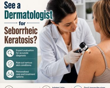
When Should You See a Dermatologist?
Schedule an appointment if you notice:
- A new dark lesion
- Rapid growth
- Persistent itching
- Frequent bleeding
- Pain
- Crusting
- Color changes
- An uncertain diagnosis
Individuals with a history of skin cancer or a strong family history should also undergo routine skin examinations.
Can Seborrheic Keratosis Become Cancerous?
One common misconception is that seborrheic keratosis eventually turns into skin cancer.
Current evidence indicates that seborrheic keratoses themselves are benign and do not become cancerous.
However, skin cancers can develop near or resemble seborrheic keratoses, which is one reason careful evaluation is important. A lesion that appears similar to seborrheic keratosis should not automatically be assumed to be harmless.
Reducing the Risk of Skin Cancer
Although seborrheic keratosis cannot always be prevented, protecting your skin from excessive ultraviolet (UV) exposure may reduce your risk of developing certain skin cancers and other sun-related skin damage.
Helpful habits include:
- Wearing broad-spectrum sunscreen with SPF 30 or higher.
- Seeking shade during peak sunlight hours.
- Wearing protective clothing and wide-brimmed hats.
- Avoiding tanning beds.
- Performing monthly skin self-examinations.
- Scheduling routine skin checks if you are at higher risk.
These measures support overall skin health and may help detect suspicious lesions earlier.
Frequently Asked Questions
Is seborrheic keratosis commonly misdiagnosed?
Yes. Because many benign and malignant skin conditions share similar features, seborrheic keratosis misdiagnosis can occur. Dermatologists sometimes use dermoscopy or a biopsy to confirm uncertain cases.
Can melanoma look exactly like seborrheic keratosis?
Yes. Some melanomas closely mimic seborrheic keratosis, making professional evaluation essential whenever a lesion appears unusual or changes over time.
Should every seborrheic keratosis be biopsied?
No. Most classic seborrheic keratoses can be diagnosed during a skin examination. A biopsy is usually reserved for lesions with atypical features or uncertain diagnoses.
Is it safe to remove a lesion at home?
No. Attempting to remove an undiagnosed skin lesion may delay diagnosis of skin cancer and increase the risk of infection or scarring. Always seek medical advice before removing a suspicious growth.
Conclusion
Seborrheic keratosis misdiagnosis is possible because these common benign skin growths often resemble other conditions, including melanoma, basal cell carcinoma, squamous cell carcinoma, actinic keratosis, and benign moles. Although most seborrheic keratoses are harmless, appearance alone is not always enough to distinguish them from potentially serious skin diseases.
A careful clinical examination, dermoscopy, and, when necessary, a skin biopsy help healthcare professionals establish an accurate diagnosis. If you notice a new skin growth, rapid changes in an existing lesion, bleeding, irregular borders, or multiple colors, seek prompt evaluation by a qualified healthcare provider. Early assessment not only provides reassurance when a lesion is benign but also supports timely detection of skin cancer when treatment is most effective.