
 By
By When you notice a rough, scaly, or crusty spot on sun-exposed skin, it can be difficult to know whether it is simply a common sun-damaged skin lesion or something more serious. Actinic keratosis vs squamous cell carcinoma is an important distinction because these two conditions can look similar, particularly in their early stages.
Actinic keratosis (AK), sometimes called solar keratosis, is considered a precancerous lesion caused primarily by long-term ultraviolet (UV) radiation exposure. Although most actinic keratoses do not become cancerous, some can progress to squamous cell carcinoma (SCC), a type of skin cancer.
What is Actinic Keratosis?
What Is Actinic Keratosis? Actinic keratosis is a common skin condition that develops after years of cumulative exposure to ultraviolet radiation. It usually appears on areas of the body that receive frequent sun exposure, such as the Actinic Keratosis on the Face, Actinic Keratosis on the Scalp, Actinic Keratosis on the Ears, Actinic Keratosis on the Neck, forearms, and backs of the hands.
An actinic keratosis may initially be easier to feel than see. The affected area can feel rough, dry, or sandpaper-like. Over time, it may become more noticeable and develop a scaly surface.
Common features include:
- Rough or scaly skin
- Dry, sandpaper-like texture
- Pink, red, brown, or skin-colored patches
- Small areas of crusting
- Mild itching, burning, or tenderness
- Lesions that persist or repeatedly return
Actinic keratoses may occur as a single spot or as multiple lesions across an area of sun-damaged skin.
Because AK represents abnormal changes in skin cells associated with chronic UV exposure, healthcare professionals often describe it as a precancerous or potentially precancerous lesion.
What Is Squamous Cell Carcinoma?
Squamous cell carcinoma is a type of skin cancer that begins in squamous cells, which are flat cells found near the surface of the skin.
SCC is commonly associated with cumulative UV exposure, although other risk factors can contribute. It may develop on sun-exposed skin but can also occur in other locations.
Squamous cell carcinoma can appear in several different ways. It may resemble a rough patch, a firm bump, a wart-like growth, or a sore that does not heal.
Some squamous cell carcinoma signs may include:
- A persistent rough or scaly patch
- A firm, raised growth
- A sore that repeatedly opens or does not heal
- Bleeding or crusting
- Increasing tenderness or pain
- A wart-like growth
- A lesion that continues to enlarge
- A rapidly changing skin lesion
Because SCC can sometimes resemble an actinic keratosis, visual appearance alone may not always provide a definite diagnosis.
Actinic Keratosis vs Squamous Cell Carcinoma: Key Differences
The main difference is that actinic keratosis represents abnormal or precancerous changes in the skin, while squamous cell carcinoma is an established skin cancer.
However, there can be significant overlap in appearance.
| Feature | Actinic Keratosis | Squamous Cell Carcinoma |
| Classification | Precancerous lesion | Skin cancer |
| Common cause | Long-term UV exposure | Often associated with UV exposure |
| Texture | Rough and scaly | Scaly, crusted, thickened, or firm |
| Appearance | Flat or slightly raised patch | May be a patch, nodule, sore, or growth |
| Pain | May be asymptomatic or mildly uncomfortable | May become tender or painful |
| Bleeding | Less typical | Can bleed or repeatedly crust |
| Growth | Often slow | May progressively enlarge |
| Diagnosis | Clinical examination; sometimes biopsy | Clinical examination and often biopsy |
| Treatment | Depends on number, location, and severity | Requires treatment by a qualified healthcare professional |
This table provides general guidance, but it should not be used to diagnose a skin lesion yourself.
Why Can Actinic Keratosis Become Squamous Cell Carcinoma?
Actinic keratosis develops because UV radiation can damage the DNA of skin cells. Repeated exposure over many years can cause abnormal cellular changes.
In some cases, these abnormal cells may continue to change and eventually develop into squamous cell carcinoma.
However, an important point is that not every actinic keratosis becomes cancer. Many remain stable, disappear, or are successfully treated before cancer develops.
The presence of multiple actinic keratoses is also a sign of significant cumulative sun damage. Therefore, people who develop AK may benefit from regular skin checks and careful sun protection.
How Do Actinic Keratosis and SCC Look Different?
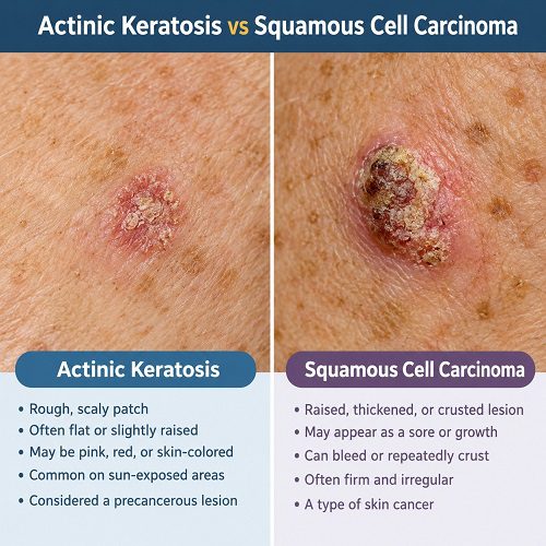
One of the challenges when comparing actinic keratosis vs squamous cell carcinoma is that both conditions can cause rough, scaly skin.
Actinic keratosis is often described as a rough patch that feels like sandpaper. It may be flat or slightly raised and can vary in color.
SCC may appear thicker, more elevated, firmer, or more irregular. It can develop into a persistent crusted area, nodule, or non-healing sore.
However, there is no universal appearance that confirms whether a lesion is benign, precancerous, or cancerous.
A lesion that changes significantly, becomes painful, bleeds, grows rapidly, or repeatedly crusts deserves professional assessment.
Squamous Cell Carcinoma Signs to Watch For
Knowing the warning signs of SCC can help you recognize when a lesion needs medical attention.
1. A sore that does not heal
A persistent sore that fails to heal within a reasonable period should be evaluated, particularly if it repeatedly bleeds or forms a crust.
2. A growing bump
A firm bump that gradually increases in size may require assessment.
3. Persistent scaling or crusting
A scaly area that continues to persist despite normal skin care should not automatically be assumed to be an actinic keratosis.
4. Bleeding
Unexpected bleeding from a skin lesion can be a warning sign, especially when the lesion repeatedly bleeds without an obvious injury.
5. Increasing pain or tenderness
An actinic keratosis may sometimes feel tender, but increasing pain or sensitivity can warrant further evaluation.
6. Rapid changes
A lesion that changes in size, shape, texture, color, or Actinic Keratosis symptoms should be checked by a healthcare professional.
Precancerous Lesions: What Does the Term Mean?
The term precancerous lesions generally refers to abnormal tissue changes that may have the potential to develop into cancer.
Actinic keratosis is one example of a precancerous skin lesion. Its presence indicates that the skin has experienced substantial sun-related damage.
Being diagnosed with a precancerous lesion does not mean that you have skin cancer. Instead, it means that monitoring and, when recommended, treatment may help reduce the risk of progression.
A healthcare professional can determine whether a lesion is likely to be an actinic keratosis or whether further investigation is needed.
Risk Factors for Actinic Keratosis
Several factors can increase the likelihood of developing actinic keratoses.
These include:
- Long-term sun exposure
- Frequent sunburns
- Outdoor occupations or activities
- Living in a region with strong sunlight
- Older age
- Fair or sun-sensitive skin
- A history of significant UV exposure
- Use of tanning beds
People with a history of substantial UV exposure may develop several lesions over time.
Risk Factors for Squamous Cell Carcinoma
Many of the risk factors for SCC overlap with those for actinic keratosis.
Potential risk factors include:
- Chronic UV exposure
- Repeated sunburn
- Previous skin cancer
- Actinic keratoses
- Weakened immune function
- Certain previous radiation exposures
- Some chronic wounds or scars
- Certain skin conditions
Having risk factors does not mean a person will develop SCC, but it can make regular skin monitoring particularly important.
How Is Actinic Keratosis Diagnosed?
A healthcare professional may diagnose actinic keratosis by examining the skin and asking about symptoms, sun exposure, and changes in the lesion.
In some cases, a biopsy may be recommended when the diagnosis is uncertain or when the lesion has features that could indicate skin cancer.
A biopsy involves removing a small sample of tissue for examination under a microscope.
This can help distinguish actinic keratosis from squamous cell carcinoma and other skin conditions.
How Is Squamous Cell Carcinoma Diagnosed?
When SCC is suspected, a healthcare professional will generally examine the lesion carefully. A skin biopsy may be required to confirm the diagnosis.
The biopsy allows a pathologist to examine the cells and determine whether cancer is present.
Because SCC can vary considerably in appearance, professional evaluation is important when a suspicious lesion develops.
Treatment Options for Actinic Keratosis
Actinic Keratosis Treatment depends on factors such as the number of lesions, their location, appearance, symptoms, and the individual’s medical history.
Common professional treatment approaches may include:
- Cryotherapy
- Prescription topical treatments
- Curettage or other procedures
- Photodynamic therapy
- Laser or other procedural approaches in selected cases
A healthcare professional can recommend the most appropriate treatment based on the individual lesion and overall skin condition.
Treatment Options for Squamous Cell Carcinoma
SCC generally requires treatment aimed at removing or destroying the cancerous cells.
Depending on the tumor’s characteristics and location, treatment may include surgical removal, Mohs micrographic surgery, curettage and electrodesiccation, radiation therapy, or other approaches for selected cases.
Early diagnosis can make treatment easier and can reduce the risk of complications.
When Should You See a Dermatologist?
You should consider professional evaluation if you notice a skin lesion that:
- Does not heal
- Keeps returning after healing
- Bleeds repeatedly
- Becomes painful or tender
- Grows quickly
- Becomes thicker or harder
- Develops significant crusting
- Changes in appearance
- Looks different from your other skin lesions
If you already have actinic keratoses, tell your healthcare professional about any lesion that looks or feels different from the others.
Can Actinic Keratosis Be Prevented?
Reducing UV exposure is one of the most important ways to protect your skin.
Helpful habits include:
- Use a broad-spectrum sunscreen as directed.
- Reapply sunscreen as recommended, especially during prolonged outdoor activity.
- Wear protective clothing when appropriate.
- Use a wide-brimmed hat for additional protection.
- Seek shade when UV levels are high.
- Avoid indoor tanning.
- Examine your skin regularly for new or changing lesions.
Sun protection remains important even after an actinic keratosis has been treated because UV damage can continue to affect surrounding skin.
Actinic Keratosis vs Squamous Cell Carcinoma: Why Early Evaluation Matters
The comparison between actinic keratosis vs squamous cell carcinoma is important because early lesions may appear similar.
Actinic keratosis is associated with sun-damaged skin and is considered a precancerous condition. SCC, on the other hand, is a form of skin cancer.
Although many actinic keratoses do not progress to cancer, some can. For this reason, persistent, changing, painful, bleeding, or rapidly growing lesions should be evaluated rather than ignored.
A healthcare professional can determine whether monitoring, treatment, or biopsy is appropriate.
Frequently Asked Questions
Is actinic keratosis the same as squamous cell carcinoma?
No. Actinic keratosis is generally considered a precancerous skin lesion associated with sun damage, while squamous cell carcinoma is a type of skin cancer. However, some actinic keratoses can progress to SCC.
Can actinic keratosis turn into squamous cell carcinoma?
Yes, some actinic keratoses can progress to SCC, although most do not. The risk varies depending on several factors, so suspicious or changing lesions should be evaluated.
What are common squamous cell carcinoma signs?
Common signs can include a persistent scaly patch, firm bump, non-healing sore, crusted lesion, bleeding, tenderness, or a growth that changes or increases in size.
Is actinic keratosis dangerous?
Actinic keratosis is not the same as skin cancer, but it indicates sun-damaged skin and has the potential to progress to SCC. Appropriate monitoring and treatment can help manage the risk.
Can you tell the difference by looking at a lesion?
Not reliably in every case. Actinic keratosis and SCC can look very similar. A dermatologist or other qualified healthcare professional may need to examine the lesion and, when necessary, perform a biopsy.
Final Thoughts
Understanding actinic keratosis vs squamous cell carcinoma can help you recognize potentially concerning skin changes. Actinic keratosis is commonly associated with long-term UV damage and is considered a precancerous lesion, while squamous cell carcinoma is a form of skin cancer.
Because the two conditions can sometimes look alike, it is not always possible to distinguish them based only on appearance. Persistent rough patches, non-healing sores, bleeding, rapid growth, increasing pain, or significant changes should be professionally evaluated.
Regular skin checks, sun protection, and timely medical assessment are important steps in protecting your long-term skin health.
Medical Disclaimer
This article is provided for general educational purposes and is not a substitute for professional medical advice, diagnosis, or treatment. If you have a new, changing, bleeding, painful, or non-healing skin lesion, consult a qualified healthcare professional or dermatologist for an appropriate evaluation.