
 By
By When you notice a new rough patch, scaly spot, or unusual bump on your skin, it can be difficult to know what it means. Some skin changes are harmless, while others may be precancerous or cancerous. Two conditions that can sometimes look similar are actinic keratosis (AK) and basal cell carcinoma (BCC).
Understanding actinic keratosis vs basal cell carcinoma is important because the two conditions are not the same. Actinic keratosis is considered a precancerous skin growth associated with long-term ultraviolet (UV) damage, while basal cell carcinoma is a type of skin cancer. Although BCC usually grows slowly and rarely spreads to distant parts of the body, it can invade surrounding tissue if left untreated.
Both conditions are strongly associated with cumulative sun exposure and commonly occur on areas such as the face, ears, scalp, neck, hands, and other sun-exposed parts of the body. However, their appearance, behavior, diagnosis, and treatment can differ. Actinic keratosis and basal cell carcinoma, including their symptoms, causes, diagnosis, treatment, and warning signs that should be evaluated by a dermatologist.
What Is Actinic Keratosis?
What Is Actinic Keratosis? Actinic keratosis is a precancerous skin growth caused primarily by long-term exposure to ultraviolet radiation from the sun or indoor tanning equipment. It develops when UV radiation damages cells in the outer layer of the skin.
Actinic keratoses commonly appear on areas that receive significant sun exposure, including:
- Actinic Keratosis on the Face
- Actinic Keratosis on the Ears
- Forehead
- Actinic Keratosis on the Scalp, especially when hair is thinning or absent
- Actinic Keratosis on the Neck
- Back of the hands
- Forearms
- Lower lip
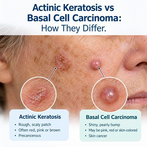
An AK may initially be easier to feel than see. The affected area can feel rough or similar to sandpaper. It may appear as a flat or slightly raised patch that is red, pink, skin-colored, gray, brown, or tan. Some lesions can become thicker or develop a horn-like appearance.
Actinic keratosis is not itself skin cancer. However, some AKs can develop into squamous cell carcinoma, another form of skin cancer. For this reason, suspicious or persistent lesions should be evaluated by a healthcare professional.
What Is Basal Cell Carcinoma?
Basal cell carcinoma is a type of skin cancer that begins in basal cells, which are found in the outer layer of the skin. It is the most common type of skin cancer and usually grows slowly. Although it rarely spreads to distant parts of the body, untreated BCC can grow deeper and damage nearby tissues.
BCC most commonly develops on areas that receive substantial sun exposure, particularly the head and neck. However, it can occur elsewhere on the body.
One of the challenges with BCC is that it does not always have a classic appearance. It may resemble a pimple, sore, scar, or irritated area. Common appearances include a shiny or pearly bump, a scaly patch, a sore that repeatedly heals and returns, or an area that looks like a scar.
Because basal cell carcinoma is cancerous, an unusual lesion that does not heal or continues changing should be assessed by a dermatologist.
Actinic Keratosis vs Basal Cell Carcinoma: Key Differences
The most important distinction is that actinic keratosis is a precancerous condition, whereas basal cell carcinoma is skin cancer.
| Feature | Actinic Keratosis | Basal Cell Carcinoma |
| Classification | Precancerous skin growth | Skin cancer |
| Main cause | Long-term UV damage | Primarily cumulative UV damage |
| Common appearance | Rough, scaly patch or spot | Shiny bump, sore, scaly patch, or scar-like lesion |
| Texture | Often rough or sandpaper-like | May be smooth, shiny, firm, crusted, or ulcerated |
| Symptoms | May itch, sting, burn, hurt, or bleed | May itch, hurt, bleed, crust, or cause sensitivity |
| Behavior | May remain stable, disappear, or progress | Usually continues to grow slowly |
| Cancer risk | Some can progress to squamous cell carcinoma | Already a form of skin cancer |
| Diagnosis | Often clinical examination; biopsy may be needed | Often requires examination and frequently biopsy for confirmation |
| Treatment | Cryotherapy, topical medicines, photodynamic therapy, or other treatments | Often surgery; other treatments may be appropriate depending on the case |
These characteristics can overlap, which is why you should not attempt to diagnose a suspicious lesion based solely on its appearance.
What Does Actinic Keratosis Look Like?
Recognizing common AK characteristics can help you know when to seek professional advice.
An actinic keratosis may appear as:
- A rough, dry, scaly patch
- A small red or pink spot
- A skin-colored or gray lesion
- A brown or tan scaly patch
- A slightly raised area
- A lesion that feels rough before it becomes visibly noticeable
- A persistent dry or scaly area on the lower lip
Some Actinic Keratosis Causes no noticeable symptoms. Others may itch, sting, burn, feel tender, or bleed.
Because AKs can resemble age spots, eczema, pimples, or other skin conditions, professional evaluation is particularly important when a lesion is new, persistent, changing, or becoming thicker.
Basal Cell Carcinoma Symptoms to Watch For
Knowing the common basal cell carcinoma symptoms can help with early detection.
A BCC may appear as:
- A shiny or pearly bump
- A firm, round, raised growth
- A pink, red, brown, black, or skin-colored lesion
- A sore that does not heal
- A sore that heals and then returns
- A scaly patch
- A growth with a depressed or indented center
- A scar-like area without an obvious injury
- A lesion that repeatedly crusts or bleeds
BCC can also cause itching, pain, numbness, tingling, or increased sensitivity in some people.
It is important to remember that basal cell carcinoma can look different from one person to another and can appear differently across skin tones. Therefore, a lesion should not be dismissed simply because it does not resemble a typical picture of skin cancer.
Why Do Actinic Keratosis and Basal Cell Carcinoma Develop?
Long-term UV exposure is an important shared risk factor.
Repeated exposure to ultraviolet radiation can damage the DNA of skin cells. Over many years, this damage can contribute to abnormal cellular changes. Actinic keratosis is one manifestation of accumulated sun damage, while basal cell carcinoma can develop when abnormal basal cells grow uncontrollably.
Risk can be influenced by factors such as:
- Long-term outdoor sun exposure
- Frequent sunburns
- Indoor tanning
- Increasing age
- A history of significant UV exposure
- Certain medical or immune-related factors
- Previous skin cancer
Importantly, people of all skin tones can develop skin cancer. Sun protection and awareness of changing skin lesions are therefore important for everyone.
Can Actinic Keratosis Turn Into Skin Cancer?
Yes, some actinic keratoses can progress to squamous cell carcinoma (SCC). This does not mean that every AK will become cancerous. However, the presence of AK indicates significant sun damage and is associated with an increased risk of developing skin cancer.
This is one reason dermatologists may recommend treatment rather than simply ignoring an AK.
It is also important to understand that AK is more directly associated with squamous cell carcinoma than basal cell carcinoma. In other words, having an AK does not mean that it will automatically turn into basal cell carcinoma.
How Are Actinic Keratosis and Basal Cell Carcinoma Diagnosed?
A dermatologist generally begins with a detailed examination of the skin. The doctor may ask when the lesion appeared, whether it has changed, and whether it causes symptoms.
Some actinic keratoses can be diagnosed through clinical examination. However, if a lesion has unusual features or there is concern about skin cancer, a biopsy may be performed.
For suspected basal cell carcinoma, a skin biopsy is commonly used to confirm the diagnosis. During a biopsy, part or all of the suspicious lesion may be removed and examined under a microscope.
This distinction is important because visual appearance alone cannot reliably determine whether a lesion is an AK, BCC, SCC, or another skin condition.
How Is Actinic Keratosis Treated?
Actinic Keratosis Treatment depends on factors such as the number of lesions, their location, appearance, thickness, and the individual’s medical circumstances.
Common treatments may include:
Cryotherapy
Liquid nitrogen can be used to freeze individual AKs. The treated area may blister, crust, and heal afterward.
Topical Treatments
For people with multiple lesions or areas of sun-damaged skin, a dermatologist may recommend prescription topical treatments designed to treat abnormal cells.
Photodynamic Therapy
Photodynamic therapy uses a photosensitizing medication followed by a specific light treatment to target abnormal cells.
Other Procedures
Depending on the lesion, location, and clinical situation, dermatologists may use other procedures to remove or destroy the abnormal tissue.
Treatment should be selected by a qualified healthcare professional rather than based solely on online photographs or home remedies.
How Is Basal Cell Carcinoma Treated?
Because BCC is skin cancer, treatment is intended to remove or destroy the cancerous cells.
Surgical treatment is commonly used. Depending on the type, location, size, and risk characteristics of the tumor, a dermatologist may recommend standard surgical excision or Mohs surgery.
Other approaches may sometimes be appropriate for selected cases, including certain topical treatments, radiation therapy, or other nonsurgical treatments. The American Academy of Dermatology notes that surgery remains a cornerstone of BCC treatment, while nonsurgical options are generally reserved for selected circumstances.
Early diagnosis is important because BCC can grow deeper into surrounding tissue even though it rarely spreads to distant organs.
When Should You See a Dermatologist?
You should consider professional evaluation if you notice a skin lesion that:
- Does not heal
- Keeps returning after healing
- Grows gradually
- Changes in color, shape, or texture
- Bleeds without an obvious reason
- Becomes painful or increasingly tender
- Develops a persistent crust
- Becomes thicker or more raised
- Looks different from surrounding skin
- Develops on an area with significant sun damage
A growing, bleeding, or changing skin spot can be an early warning sign of skin cancer.
Do not wait for a lesion to become painful before seeking advice. Some basal cell carcinomas cause few or no symptoms during their early stages.
How Can You Reduce Your Risk?
Because UV exposure plays an important role in both actinic keratosis and many cases of skin cancer, protecting your skin is an important part of prevention.
Consider the following habits:
- Use a broad-spectrum sunscreen regularly.
- Reapply sunscreen as directed, especially during prolonged outdoor activities.
- Wear protective clothing when spending time outdoors.
- Use a wide-brimmed hat when appropriate.
- Seek shade during periods of strong sunlight.
- Avoid indoor tanning beds and sunlamps.
- Regularly check your skin for new or changing lesions.
- Schedule professional skin examinations when recommended.
For people who have already developed actinic keratoses, sun protection is particularly important because significant UV damage increases the risk of additional AKs and skin cancer.
Actinic Keratosis vs Basal Cell Carcinoma: What Should You Remember?
The key difference between actinic keratosis vs basal cell carcinoma is their classification and behavior.
Actinic keratosis is a precancerous growth caused by cumulative UV damage. It often appears as a rough, scaly patch on sun-exposed skin and may cause itching, burning, tenderness, or no symptoms at all.
Basal cell carcinoma, on the other hand, is a form of skin cancer. It commonly appears as a shiny or pearly bump, persistent sore, scaly patch, or scar-like growth. It usually grows slowly and rarely spreads, but it can damage nearby tissue when left untreated.
The two conditions can sometimes look alike, and photographs cannot provide a reliable diagnosis. If you notice a new, persistent, changing, bleeding, or non-healing skin lesion, a dermatologist can determine whether it requires monitoring, treatment, or further testing.
Final Thoughts
Learning the differences between actinic keratosis and basal cell carcinoma can help you become more aware of changes in your skin. An actinic keratosis is not the same as skin cancer, but it is a sign of significant UV-related skin damage and can sometimes progress to squamous cell carcinoma.
Basal cell carcinoma is already a form of skin cancer and should be diagnosed and treated appropriately. Fortunately, early detection and treatment of BCC generally lead to very good outcomes.
If you are unsure whether a rough patch, bump, sore, or changing spot is an actinic keratosis, basal cell carcinoma, or another skin condition, avoid self-diagnosing. A dermatologist can examine the lesion and determine whether additional testing, such as a biopsy, is needed.
Medical Disclaimer
This article is for educational and informational purposes only and does not provide medical advice, diagnosis, or treatment. If you have a new, changing, bleeding, painful, or non-healing skin lesion, consult a qualified dermatologist or healthcare professional for appropriate evaluation.