 By
By Actinic keratosis and seborrheic keratosis are two common types of skin lesions that can look similar at first glance. Both may appear as rough, scaly or raised patches on the skin, particularly as people get older. However, they have very different causes, risks and treatment considerations.
Understanding actinic keratosis vs seborrheic keratosis is important because actinic keratosis can sometimes develop into squamous cell carcinoma, a type of skin cancer. Seborrheic keratosis, on the other hand, is generally harmless and does not turn into skin cancer.
Although appearance can provide useful clues, it is not always possible to distinguish these conditions accurately without a professional skin examination. A dermatologist may use dermoscopy or recommend a biopsy when a lesion has unusual features. The differences between these two common skin growths, including their causes, appearance, symptoms, risk factors, diagnosis and treatment.
What is Actinic Keratosis?
What Is Actinic Keratosis? Actinic keratosis (AK), also called solar keratosis, is a precancerous skin lesion caused primarily by long-term exposure to ultraviolet (UV) radiation.
It develops when UV radiation damages skin cells over many years. As a result, abnormal cells begin to grow in the outer layer of the skin.
Actinic keratoses are particularly common in areas that receive frequent sun exposure, including:
- Actinic Keratosis on the Face
- Actinic Keratosis on the Scalp, especially in people with thinning or no hair
- Actinic Keratosis on the Ears
- Actinic Keratosis on the Neck
- Forearms
- Actinic Keratosis on the Hands
- Lower legs in some people
The lesions are usually small, but they may become larger over time or occur in groups.
Actinic keratosis is considered a precancerous condition because some lesions can progress to squamous cell carcinoma (SCC). Not every actinic keratosis becomes cancerous, but identifying and treating suspicious lesions can help reduce potential risks.
What Is Seborrheic Keratosis?
Seborrheic keratosis is a common non-cancerous skin growth. It usually develops with age and may appear on the chest, back, abdomen, scalp, face or other areas of the body.
Unlike actinic keratosis, seborrheic keratosis is not considered a precancerous lesion.
These growths can vary considerably in appearance. They may be:
- Light tan
- Brown
- Dark brown
- Black
- Pinkish or skin-coloured
They often have a waxy, rough or wart-like surface. One of their classic characteristics is that they can appear as though they have been “stuck on” the surface of the skin.
Seborrheic keratoses may occur as a single growth or multiple lesions. Some people develop many of them as they age.
Actinic Keratosis vs Seborrheic Keratosis: Key Differences
While the two conditions can overlap in appearance, several characteristics can help distinguish them.
| Feature | Actinic Keratosis | Seborrheic Keratosis |
| Main cause | Long-term UV exposure | Age-related skin growth |
| Cancer risk | Precancerous; may progress to SCC | Generally benign |
| Texture | Often rough and scaly | Waxy, rough or wart-like |
| Typical colour | Pink, red, tan or skin-coloured | Tan, brown, dark brown, black or skin-coloured |
| Common locations | Sun-exposed skin | Trunk, face, scalp and other areas |
| Pain/tenderness | May be tender or sensitive | Usually painless |
| Typical appearance | Flat or slightly raised scaly patch | Raised, waxy or “stuck-on” growth |
| Treatment | Often treated to prevent progression | Usually treated only if troublesome or uncertain |
| UV relationship | Strong association | Not primarily caused by UV exposure |
The table provides general clues, but individual lesions can look different. A dermatologist should assess lesions that are new, changing or unusual.
Differences in Appearance
Appearance is one of the most useful clues when comparing seborrheic keratosis vs Actinic Keratosis, although visual diagnosis alone is not always reliable.
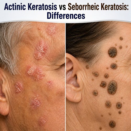
Actinic Keratosis Appearance
Actinic keratosis often begins as a small, rough patch of skin. It may feel easier to identify by touch than by sight.
The affected area can feel:
- Dry
- Rough
- Scaly
- Sandpaper-like
- Slightly thickened
The colour may range from skin-coloured or pale pink to red, brown or grey.
Some lesions are barely visible but can be felt when you run a finger over the skin. They may gradually become thicker or develop a more noticeable scale.
Seborrheic Keratosis Appearance
Seborrheic keratoses are often more clearly raised than actinic keratoses. They may have a waxy, greasy or wart-like appearance.
A seborrheic keratosis may look as though it has been placed on top of the skin rather than growing from within it.
They can range from very small spots to larger growths. Some are smooth, while others have a rough, irregular surface.
Because seborrheic keratoses can sometimes resemble skin cancer, an unusual lesion should be examined rather than assumed to be benign.
Causes and Risk Factors
The causes of the two conditions are another important difference.
Causes of Actinic Keratosis
The primary cause of actinic keratosis is cumulative UV radiation.
Risk increases with:
- Frequent sun exposure
- Outdoor work or activities
- Repeated sunburn
- Older age
- Fair skin
- A history of significant UV exposure
- Use of tanning beds
The damage responsible for actinic keratosis accumulates over many years. This means lesions may appear long after the original UV exposure occurred.
Causes of Seborrheic Keratosis
The exact cause of seborrheic keratosis is not completely understood. However, these growths become increasingly common with age.
Genetic factors may also play a role, particularly in people who develop multiple lesions.
Unlike actinic keratosis, seborrheic keratosis is not regarded as a direct result of cumulative UV damage.
Symptoms of Actinic Keratosis
Actinic Keratosis Symptoms may vary, and many actinic keratoses do not cause significant symptoms. However, some people may experience:
- Rough or scaly skin
- Mild itching
- Burning
- Tenderness
- Sensitivity
- Occasional bleeding or irritation
A lesion may feel rough before it becomes visibly obvious.
A particularly important warning sign is a lesion that becomes thicker, painful, rapidly changes or develops bleeding or ulceration. These features warrant medical assessment because they can sometimes indicate progression towards squamous cell carcinoma.
Symptoms of Seborrheic Keratosis
Seborrheic keratoses are usually asymptomatic.
However, they can sometimes:
- Itch
- Become irritated by clothing
- Catch on jewellery
- Bleed after scratching or rubbing
- Become inflamed
Because these growths can become irritated, some people choose to have them removed even though they are generally harmless.
A sudden change in an existing growth should still be assessed, particularly if the diagnosis has never been confirmed.
Where Do These Lesions Usually Appear?
Location can provide another clue.
Actinic keratosis is strongly associated with areas exposed to the sun. Common sites include the face, ears, scalp, neck, hands and forearms.
Seborrheic keratosis can develop almost anywhere except the palms and soles. It is especially common on the chest, back, abdomen, scalp and face.
However, location alone cannot confirm the diagnosis. Both conditions can occasionally occur in areas that do not fit their most typical patterns.
Can Actinic Keratosis Turn Into Skin Cancer?
Yes. Actinic keratosis is considered a precancerous lesion because some lesions can develop into squamous cell carcinoma.
The overall risk for an individual lesion is relatively low, but people with multiple actinic keratoses may have evidence of substantial cumulative UV damage and may have an increased risk of developing skin cancer.
This is why actinic keratoses should not simply be ignored.
Regular skin checks and appropriate treatment can help manage existing lesions and identify suspicious changes early.
Can Seborrheic Keratosis Become Cancerous?
Seborrheic keratosis is generally benign and does not normally transform into skin cancer.
However, the challenge is that some skin cancers can resemble seborrheic keratoses.
For example, melanoma, basal cell carcinoma and squamous cell carcinoma can occasionally have a similar appearance to a benign growth.
Therefore, if a lesion looks unusual or changes unexpectedly, a dermatologist may recommend further examination or biopsy.
How Are They Diagnosed?
A doctor or dermatologist will usually begin by examining the lesion and asking about its history.
Important questions may include:
- When did the lesion first appear?
- Has it changed in size or colour?
- Does it itch or hurt?
- Has it bled?
- Is it becoming thicker?
- Have you had significant sun exposure?
- Do you have a history of skin cancer?
Dermoscopy may be used to examine structures beneath the surface of the skin.
If the appearance is uncertain, a biopsy may be recommended. During a biopsy, a small portion or the entire lesion may be removed and examined under a microscope.
This can help determine whether the lesion is actinic keratosis, seborrheic keratosis or another skin condition.
Treatment for Actinic Keratosis
Actinic Keratosis Treatment depends on factors such as the number of lesions, their location, thickness and appearance.
Common treatment options include:
Cryotherapy
Cryotherapy uses liquid nitrogen to freeze individual lesions. The treated area may blister, crust and heal over the following days or weeks.
Prescription Creams
Certain topical medicines can treat visible actinic keratoses as well as areas of surrounding sun-damaged skin.
Depending on the circumstances, doctors may prescribe treatments such as 5-fluorouracil, imiquimod or other appropriate therapies.
Photodynamic Therapy
Photodynamic therapy uses a photosensitising medication followed by exposure to a specific type of light. It can be useful when there are multiple actinic keratoses in an area.
Curettage or Other Procedures
Thicker or suspicious lesions may require scraping, removal or another procedure. If there is concern about skin cancer, the lesion may be biopsied or completely removed.
Treatment for Seborrheic Keratosis
Seborrheic keratoses usually do not require treatment.
Removal may be considered if the growth:
- Becomes irritated
- Bleeds repeatedly
- Catches on clothing
- Causes discomfort
- Is cosmetically bothersome
- Has an uncertain diagnosis
Possible removal methods include cryotherapy, curettage or other dermatological procedures.
The appropriate method depends on the size, thickness, location and appearance of the growth.
When Should You See a Dermatologist?
It can be difficult to distinguish between different rough skin lesions without professional assessment.
Seek medical advice if a skin lesion:
- Changes rapidly
- Becomes significantly thicker
- Bleeds without an obvious reason
- Develops an ulcer
- Becomes persistently painful
- Has an irregular or unusual appearance
- Develops a new colour
- Does not heal
- Looks noticeably different from your other lesions
The “ugly duckling” principle can also be useful: a lesion that looks noticeably different from the other marks or growths on your skin deserves attention.
Prevention and Skin Protection
Although seborrheic keratosis cannot necessarily be prevented, protecting your skin from UV radiation is important for reducing the development of actinic keratoses and lowering your overall risk of skin cancer.
Consider the following habits:
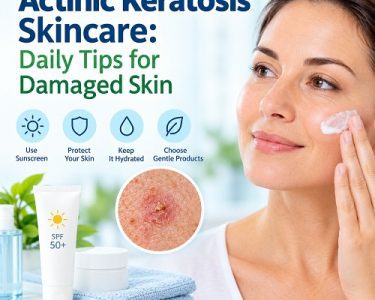
- Use a broad-spectrum sunscreen with an appropriate SPF.
- Reapply sunscreen as directed, particularly when outdoors.
- Wear protective clothing.
- Use a wide-brimmed hat when exposed to strong sunlight.
- Seek shade during periods of intense UV exposure.
- Avoid tanning beds.
- Check your skin regularly for new or changing lesions.
Sun protection is particularly important for people who have already developed actinic keratoses.
Actinic Keratosis vs Seborrheic Keratosis: What Is the Main Difference?
The biggest distinction is their potential health risk.
Actinic keratosis is associated with long-term UV damage and is considered precancerous. Some lesions can progress to squamous cell carcinoma, so monitoring and treatment may be recommended.
Seborrheic keratosis is generally a harmless, non-cancerous skin growth. It may be removed if it becomes irritated or if there is uncertainty about the diagnosis.
Both conditions can be rough, scaly or pigmented, which is why it is sometimes difficult to tell them apart based only on appearance.
If you have a new or changing skin growth, it is better to have it professionally assessed rather than attempting to diagnose it yourself.
Final Thoughts
Understanding actinic keratosis vs seborrheic keratosis can help you recognise why apparently similar skin lesions may require very different approaches.
Actinic keratosis is usually linked to cumulative sun damage and may appear as a rough, scaly patch on sun-exposed skin. Because it is considered precancerous, appropriate evaluation and treatment are important.
Seborrheic keratosis is generally benign and commonly develops with age. It may look waxy, raised or “stuck on” to the skin and usually does not require treatment unless it causes problems or the diagnosis is uncertain.
Most importantly, not every unusual skin growth is harmless simply because it resembles a seborrheic keratosis. A dermatologist can assess suspicious lesions and determine whether further testing is necessary.
Early evaluation is particularly important when a lesion changes rapidly, bleeds, becomes painful, thickens or looks different from your other skin growths.
Medical Disclaimer
This article is intended for general educational purposes only and should not replace professional medical advice, diagnosis or treatment. If you have a new, changing, bleeding or suspicious skin lesion, consult a qualified dermatologist or healthcare professional for an appropriate examination.