 By
By When you notice a new dark, raised, or unusual spot on your skin, it is natural to wonder what it might be. Two common possibilities are seborrheic keratosis and moles. Although these skin growths can sometimes look similar, they are different conditions with distinct characteristics.
Understanding seborrheic keratosis vs mole can help you recognize common differences in appearance, texture, location, and development. However, because some skin lesions can resemble one another—and because certain skin cancers can sometimes look like benign growths—an unusual or changing spot should be evaluated by a qualified healthcare professional.
What Is Seborrheic Keratosis?
Seborrheic keratosis is a common noncancerous skin growth. It often appears as a raised, rough, waxy, or “stuck-on” growth on the skin. These growths become more common with age and may occur on the chest, back, abdomen, shoulders, scalp, or other areas.
Seborrheic keratoses can vary considerably in appearance. Some are light tan or beige, while others may be brown, dark brown, or nearly black. Their surfaces can be smooth, rough, flaky, or waxy.
One of their most recognizable features is their appearance as though they have been attached to the surface of the skin. They may also become irritated by clothing, jewelry, or scratching.
Seborrheic keratoses are generally harmless and do not usually require treatment. If one becomes irritated, bleeds after minor trauma, changes noticeably, or looks unusual, however, a dermatologist may recommend an examination.
What Is a Mole?
A mole, medically known as a nevus, is a common type of skin growth that develops from melanocytes, the cells responsible for producing melanin, the pigment that gives skin its color.
Moles can be flat or raised and may range from pink or tan to brown or black. Many people have multiple moles, and they can appear on almost any part of the body.
Most ordinary moles are harmless. Some are present from birth, while others develop during childhood, adolescence, or adulthood.
Moles can change over time because of normal aging, hormonal changes, or other factors. Nevertheless, a mole that develops significant changes should be assessed by a healthcare professional because certain melanoma—the most serious form of skin cancer—can develop in or resemble a mole.
Seborrheic Keratosis vs Mole: Key Differences
When comparing seborrheic keratosis vs mole, several characteristics can provide useful clues.
1. Surface and Texture
Texture is often one of the easiest differences to notice.
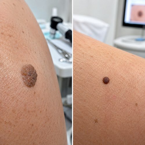
Seborrheic keratoses commonly have a rough, waxy, scaly, or crust-like surface. They may feel elevated and textured when touched.
Moles can be raised, but many are relatively smooth. A mole may feel like part of the surrounding skin rather than having a waxy or “stuck-on” texture.
However, texture alone cannot establish a diagnosis.
2. Overall Appearance
Seborrheic keratoses often look as though something has been placed on top of the skin. Their edges may be sharply defined, and their surface can appear waxy.
Moles are usually more integrated with the surrounding skin. They may be flat, slightly raised, or dome-shaped.
Because both can be brown or darkly pigmented, color alone is not enough to distinguish them.
3. Color
Seborrheic keratoses can range from pale tan to dark brown or almost black. Some may contain several shades within the same growth.
Moles may also be tan, brown, dark brown, black, pink, or skin-colored.
A new lesion with Multiple Seborrheic Keratosis unusual colors, especially when accompanied by other concerning changes, deserves professional evaluation.
4. Shape
Seborrheic keratoses often have a round or oval appearance, although their shapes can vary.
Common moles are frequently round or oval and may have relatively regular borders.
An irregularly shaped lesion is not automatically cancerous, but a noticeable change in shape can be a reason to seek medical advice.
5. Development Over Time
Seborrheic keratoses often develop gradually and may increase in number as people get older.
Moles can develop at different stages of life. Some appear during childhood or adolescence, while others develop later.
A new growth in adulthood is not necessarily dangerous, but it should be monitored, particularly if it looks unusual or changes rapidly.
Can Seborrheic Keratosis Look Like a Mole?
Yes. This is one reason people may have difficulty identifying the difference between seborrheic keratosis vs mole.
A darker seborrheic keratosis can resemble a dark mole, particularly when viewed without magnification. Some seborrheic keratoses are relatively flat, while others can become significantly raised.
Likewise, some moles can be raised and pigmented, making them resemble seborrheic keratoses.
A dermatologist may use dermoscopy, a specialized skin examination technique, to look at structures that are not easily visible with the naked eye. In uncertain cases, a biopsy may be recommended to determine exactly what a lesion is.
Seborrheic Keratosis vs Mole: Where They Appear
Both seborrheic keratoses and moles can occur on many parts of the body.
Seborrheic keratoses commonly appear on the:
- Chest
- Back
- Abdomen
- Shoulders
- Neck
- Scalp
- Face
Moles can also appear on the face, neck, arms, legs, trunk, and other areas.
Therefore, location alone usually cannot tell you whether a growth is a seborrheic keratosis or a mole.
Are Seborrheic Keratoses Dangerous?
Seborrheic keratoses are generally benign. They are not considered skin cancer and typically do not become melanoma.
However, some lesions can look unusual or resemble other skin conditions. A growth that bleeds, becomes inflamed, changes rapidly, or develops an unfamiliar appearance should not automatically be assumed to be a seborrheic keratosis.
It is particularly important not to self-diagnose a dark or changing skin growth based only on photographs or online descriptions.
Are Moles Dangerous?
Most moles are harmless. However, some moles may have features that require closer monitoring.
Melanoma can sometimes develop from an existing mole, although many melanomas arise on previously normal-looking skin. This is why paying attention to changes in your skin is important.
A useful approach is to become familiar with the appearance of your existing moles and look for noticeable changes.
Warning Signs to Watch For
The ABCDE method is commonly used as a reminder of potential warning signs associated with melanoma:
A – Asymmetry: One half of the lesion looks different from the other half.
B – Border: The edges are irregular, poorly defined, scalloped, or uneven.
C – Color: The lesion contains multiple or unusual colors.
D – Diameter: The lesion is larger than about 6 millimeters, although melanoma can be smaller.
E – Evolving: The lesion changes in size, shape, color, elevation, or symptoms.
Other concerning symptoms may include persistent itching, tenderness, crusting, or bleeding.
These signs do not automatically mean that a lesion is melanoma. They simply indicate that professional evaluation may be appropriate.
When Should You See a Dermatologist?
Consider arranging a professional skin examination if you notice a growth that:
- Is new and unusual
- Changes significantly
- Becomes darker or develops several colors
- Has an irregular border
- Bleeds without obvious injury
- Develops persistent crusting
- Becomes painful or persistently itchy
- Looks substantially different from your other skin growths
- You cannot confidently identify
A dermatologist can examine the lesion and determine whether it is consistent with seborrheic keratosis, a mole, another benign condition, or something requiring further investigation.
Can Seborrheic Keratosis Be Removed?
Seborrheic keratoses usually do not need treatment when they are clearly benign and are not causing problems.
Removal may be considered when a growth is Irritated Seborrheic Keratosis, repeatedly catches on clothing, becomes bothersome, or for cosmetic reasons.
Common professional removal methods include Cryotherapy for Seborrheic Keratosis, curettage, and other dermatologic techniques. The appropriate method depends on the growth’s size, location, thickness, and appearance.
It is best to avoid attempting to cut, burn, scrape, or chemically remove a skin growth at home. Such methods can Seborrheic Keratosis Causes infection, scarring, bleeding, and may make it harder for a professional to evaluate the lesion.
Can Moles Be Removed?
Moles can also be removed by a qualified healthcare professional when medically appropriate or when they are bothersome or cosmetically unwanted.
Depending on the mole and clinical circumstances, a dermatologist may use a procedure such as surgical excision or another appropriate technique.
If a mole appears suspicious, the removed tissue may be examined under a microscope to determine whether abnormal or cancerous cells are present.
Natural Skin Care and Seborrheic Keratosis
People searching for information about seborrheic keratosis vs mole may also encounter home remedies and natural approaches online.
Gentle skin care for Seborrheic Keratosis can help maintain the skin barrier, but there is no reliable home remedy that can safely diagnose or remove a suspicious skin growth.
Avoid applying strong acids, essential oils, abrasive substances, or other unproven products directly to a pigmented growth. These approaches can irritate the skin and potentially cause burns or scarring.
If you are interested in natural skin care, focus on gentle cleansing, regular moisturizing, sun protection, and avoiding unnecessary irritation.
Natural Remedies for Seborrheic Keratosis
Seborrheic keratosis is a common, noncancerous skin growth that often develops with age. Although these growths are usually harmless, they can sometimes become irritated, itchy, or uncomfortable. Many people look for Natural Remedies for Seborrheic Keratosis as part of a gentle approach to skin care.
It is important to understand that there is no scientifically proven natural remedy that safely removes seborrheic keratosis. Instead, Natural Support for Seborrheic Keratosis should focus on keeping the surrounding skin healthy and reducing irritation. Gentle cleansing, regular moisturizing, and avoiding scratching or picking can help protect the skin.
Seborrheic Keratosis in Older Adults is particularly common, so older individuals may benefit from regular skin checks and good daily skin-care habits. Protecting the skin from excessive sun exposure with appropriate sunscreen and protective clothing is also a useful part of overall skin health.
For Seborrheic Keratosis Home Care, avoid harsh acids, abrasive scrubs, essential oils, or attempts to cut or burn the growth. These methods may cause irritation, infection, or scarring.
If a growth changes rapidly, bleeds, becomes painful, or looks unusual, consult a dermatologist. Professional evaluation is especially important because some other skin conditions can resemble seborrheic keratosis. A healthcare professional can confirm the diagnosis and discuss safe treatment options when necessary.
How Doctors Tell the Difference
When a dermatologist evaluates a skin growth, they consider several factors rather than relying on a single characteristic.
The examination may include the lesion’s:
- Color
- Shape
- Border
- Texture
- Size
- Location
- Pattern
- Development over time
- Changes compared with previous examinations
Dermoscopy can provide a closer view of structures within the lesion. If the diagnosis remains uncertain, a biopsy may provide a definitive answer.
This is especially important when a lesion could potentially resemble melanoma or another skin condition.
Seborrheic Keratosis vs Mole: A Simple Comparison
| Feature | Seborrheic Keratosis | Mole |
| Typical nature | Benign skin growth | Usually benign nevus |
| Texture | Often rough, waxy, or scaly | Often smooth, but may be raised |
| Appearance | May look “stuck on” | Usually integrated with skin |
| Color | Tan, brown, dark brown, or black | Skin-colored, tan, brown, pink, or black |
| Shape | Round, oval, or irregular | Often round or oval |
| Development | More common with increasing age | Often develops during childhood or adulthood |
| Cancer risk | Generally benign | Most are benign, but some lesions can be associated with melanoma |
| Evaluation | Needed when unusual or changing | Important when changing or suspicious |
The Bottom Line
Understanding seborrheic keratosis vs mole can help you become more familiar with common skin growths, but appearance alone cannot always provide a reliable diagnosis.
Seborrheic keratoses are generally benign growths that often have a rough, waxy, or stuck-on appearance. Moles are collections of pigment-producing cells and can be flat or raised, smooth or slightly textured, and vary in color.
The most important difference is not simply whether a spot is brown, black, raised, or flat. What matters is how the lesion looks overall and whether it is changing.
If you notice a new, unusual, rapidly changing, bleeding, painful, or otherwise concerning skin growth, arrange an examination with a dermatologist. Professional evaluation is the safest way to determine what a suspicious lesion actually is.
Frequently Asked Questions
Can a seborrheic keratosis be mistaken for a mole?
Yes. Dark or raised seborrheic keratoses can sometimes resemble moles. A dermatologist can use a clinical examination and, when needed, dermoscopy or biopsy to clarify the diagnosis.
Are seborrheic keratoses cancerous?
Seborrheic keratoses are generally benign and are not considered skin cancer. However, an unusual growth should still be evaluated rather than automatically assumed to be seborrheic keratosis.
Can a mole turn into melanoma?
Some melanomas can develop in existing moles, while others develop independently. Any mole that changes noticeably should be evaluated by a healthcare professional.
What is the biggest difference between a seborrheic keratosis and a mole?
A seborrheic keratosis often has a waxy, rough, scaly, or stuck-on appearance. Moles are commonly smoother and may be flat or raised. However, there can be significant overlap in appearance.
Should I remove a seborrheic keratosis at home?
Home removal is not recommended. Cutting, burning, scraping, or applying harsh chemicals can cause injury, infection, and scarring. Professional assessment is safer, especially when the diagnosis is uncertain.