 By
By A new or unusual skin growth can be concerning, especially when it is dark, raised, rough, or changes over time. Two conditions that can sometimes look similar are seborrheic keratosis vs melanoma. Although seborrheic keratoses are usually harmless, melanoma is a type of skin cancer that can become serious if it spreads.
Knowing the difference can help you recognize when a skin lesion may need professional evaluation. However, appearance alone cannot always provide a definite diagnosis. Some melanomas can resemble common moles, age spots, cysts, or other harmless growths. A dermatologist may need to examine the lesion with specialized equipment and, when necessary, remove it for microscopic examination.
What is Seborrheic Keratosis?
Seborrheic keratosis is a common, noncancerous skin growth. It becomes more common during middle age and later in life. These growths can appear on areas such as the chest, back, neck, scalp, and face.
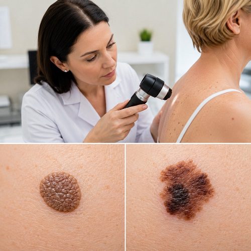
A typical seborrheic keratosis often has a waxy, rough, or “stuck-on” appearance. It may begin as a small rough bump and gradually become thicker. Colors can vary from light tan and brown to very dark brown or black.
Most seborrheic keratoses do not cause serious problems and do not require treatment. Some may itch or become irritated when they rub against clothing, jewelry, or other surfaces.
Despite being harmless, seborrheic keratoses can sometimes look concerning. A dark or irregular growth may be difficult for someone without medical training to distinguish from melanoma.
What Is Melanoma?
Melanoma is a type of skin cancer that develops from pigment-producing cells called melanocytes. It is considered one of the more serious forms of skin cancer because it can spread to other parts of the body. When detected early, however, melanoma is highly treatable.
Melanoma may appear as a new spot or develop within an existing mole. It does not always look like the classic dark, black mole people associate with skin cancer. Some melanomas can contain shades of brown, black, red, pink, white, or blue.
This is why changes in a skin lesion are often more important than simply looking at its color.
Seborrheic Keratosis vs Melanoma: Key Differences
When comparing seborrheic keratosis vs melanoma, several features can provide useful clues.
1. Overall Appearance
Seborrheic keratoses often have a raised, waxy, rough, or wart-like appearance. They may look as though they have been placed or “stuck” onto the skin.
Melanoma can have a flatter or raised appearance and may resemble a mole, freckle, age spot, or other skin growth.
Because melanoma can have different appearances, there is no single visual feature that can reliably rule it out.
2. Shape
A typical seborrheic keratosis may have a fairly recognizable shape and appearance.
Melanoma may be asymmetrical, meaning one half does not resemble the other half. Asymmetry is the first warning sign in the commonly used ABCDE method for identifying suspicious pigmented lesions.
3. Border
Seborrheic keratoses may have a relatively well-defined appearance, although their edges can vary.
A melanoma may have an irregular, scalloped, notched, or poorly defined border. An uneven border should therefore be considered a reason to have a suspicious lesion examined.
4. Color
Seborrheic keratoses can Multiple Seborrheic Keratosis from white to black, although tan and brown shades are common.
Melanomas may contain multiple colors or shades within the same lesion. These can include different shades of brown or black as well as areas of red, white, or blue.
However, color alone does not determine whether a lesion is cancerous. A very dark seborrheic keratosis can still be benign, while some melanomas may have little pigment.
5. Growth and Change
One of the most important differences in seborrheic keratosis vs melanoma is the way a lesion changes.
Seborrheic keratoses often develop gradually and may become thicker over time. Melanoma may change in size, shape, color, or appearance.
The “E” in the ABCDE system stands for Evolving, meaning a spot is changing in a noticeable way.
A new or changing lesion deserves attention, particularly when the change is rapid or accompanied by other warning signs.
The ABCDE Warning Signs of Melanoma
The ABCDE method is one of the most useful ways to remember potential melanoma warning signs.
A — Asymmetry
Imagine dividing a spot into two halves. If one half looks significantly different from the other, the lesion may be suspicious.
B — Border
Look for borders that are irregular, jagged, scalloped, notched, or poorly defined.
C — Color
Be cautious when a lesion contains multiple colors or noticeable variations in pigmentation. These may include brown, black, tan, red, white, or blue areas.
D — Diameter
Melanomas are often larger than 6 millimeters when diagnosed, approximately the size of a pencil eraser. However, melanoma can be smaller, so size alone should never be used to rule it out.
E — Evolving
A spot that changes in size, shape, color, or overall appearance deserves medical attention.
The American Academy of Dermatology recommends seeing a dermatologist if you notice a new spot, a spot that looks different from your other lesions, or a lesion that changes, itches, or bleeds.
Warning Signs You Should Never Ignore
Not every unusual skin growth is melanoma, but certain changes should not be ignored.
A Skin Growth That Changes Quickly
Rapid changes in a lesion can be concerning. If a growth suddenly becomes larger, darker, more irregular, or noticeably different, arrange an evaluation with a dermatologist.
A Lesion That Bleeds
Seborrheic keratoses can occasionally become Irritated Seborrheic Keratosis and bleed, particularly when rubbed or scratched. Nevertheless, unexplained or repeated bleeding should be evaluated rather than automatically attributed to irritation.
The American Academy of Dermatology specifically advises seeing a dermatologist when a seborrheic keratosis grows quickly, turns black, itches, bleeds, or differs from the typical appearance of a seborrheic keratosis.
Persistent Itching or Pain
Many seborrheic keratoses cause no symptoms, although some may itch. Melanoma can also sometimes itch or become painful, although it may cause no discomfort at all.
Therefore, Seborrheic Keratosis symptoms should be considered alongside changes in appearance.
A New or Unusual Spot
A new lesion that looks different from your other spots can be important. Dermatologists sometimes describe this as the “ugly duckling” concept: a lesion that stands out from the rest may deserve closer examination.
A Sore That Does Not Heal
A spot that repeatedly breaks down, bleeds, heals, and returns should not be ignored. Persistent or unusual skin changes deserve professional evaluation.
A Dark Line Under a Nail
Melanoma can also develop under a fingernail or toenail. A new or changing dark line beneath a nail should be assessed by a healthcare professional, especially if it changes or becomes wider.
Can Seborrheic Keratosis Turn Into Melanoma?
Seborrheic keratosis itself is considered a benign, noncancerous growth. It is not generally regarded as a precursor to melanoma.
The concern is that a melanoma can sometimes look like a seborrheic keratosis. This can make visual identification challenging.
For this reason, do not assume that every dark, rough, or raised growth is simply an age-related seborrheic keratosis. If a lesion looks unusual or changes over time, it should be examined.
Why Self-Diagnosis Can Be Difficult
Searching online for pictures of seborrheic keratosis and melanoma can be helpful for learning general warning signs, but photographs cannot provide a reliable diagnosis for an individual lesion.
Skin lesions can vary significantly from person to person. Lighting, skin tone, lesion size, location, and stage can also affect appearance.
A dermatologist can use a dermatoscope to examine a suspicious lesion more closely. If the diagnosis remains uncertain or skin cancer is suspected, the lesion may be removed or sampled and examined under a microscope. This microscopic examination is what can establish whether cancer cells are present.
How Dermatologists Evaluate a Suspicious Growth
During a skin examination, a dermatologist may ask when you first noticed the lesion and whether it has changed.
The doctor may assess:
- Shape
- Size
- Color
- Border
- Surface texture
- Location
- Recent changes
- Bleeding or itching
- Whether it looks different from your other skin lesions
A dermatoscope may provide a magnified view of structures that are difficult to see with the naked eye.
If a lesion appears suspicious, the dermatologist may recommend a biopsy or removal. The tissue is then examined under a microscope to determine the diagnosis.
How to Check Your Skin at Home
Regular skin self-examination can help you notice changes early.
Use a well-lit room and a full-length mirror. A handheld mirror can help you examine areas that are difficult to see.
Check your:
- Face and neck
- Chest and abdomen
- Arms and hands
- Legs and feet
- Back
- Scalp
- Palms and soles
- Areas between the toes
- Fingernails and toenails
When checking your skin, pay particular attention to new spots or existing lesions that have changed.
You can also take photographs of suspicious-looking areas for personal comparison over time. However, photographs should complement—not replace—a professional examination when a lesion is concerning.
Who Should Be Especially Careful?
Anyone can develop melanoma, regardless of skin color.
Your personal risk may be higher if you have factors such as a personal history of melanoma, significant ultraviolet exposure, or other risk factors identified by your healthcare professional.
People with numerous moles or a family history of melanoma may also benefit from discussing appropriate skin-monitoring strategies with a dermatologist.
The important point is not to assume that skin cancer only affects people with a particular skin tone or age.
When Should You See a Dermatologist?
Consider arranging a professional skin examination if you notice a lesion that:
- Is new and unusual
- Changes in size, shape, or color
- Has an irregular border
- Contains several different colors
- Becomes increasingly dark
- Grows quickly
- Bleeds without an obvious reason
- Repeatedly becomes irritated
- Itches or hurts persistently
- Looks noticeably different from your other spots
- Does not heal normally
The American Academy of Dermatology advises prompt dermatological assessment for spots that are changing, itching, bleeding, or otherwise suspicious.
This does not mean that every such lesion is melanoma. It simply means that the cause should be determined by someone qualified to evaluate skin abnormalities.
Should You Remove a Seborrheic Keratosis at Home?
No. Trying to cut, burn, scrape, or chemically remove a skin growth yourself can cause bleeding, infection, scarring, and delayed diagnosis.
If a lesion thought to be a seborrheic keratosis looks unusual, a dermatologist may recommend removing it so that it can be examined under a microscope.
Professional assessment is particularly important when there is uncertainty about whether a growth is benign.
Natural Support for Seborrheic Keratosis
Natural Support for Seborrheic Keratosis can focus on keeping the surrounding skin comfortable, moisturized, and protected. Seborrheic keratoses are common, noncancerous skin growths that may appear rough, raised, waxy, or brown. Although they usually do not require treatment, irritated growths can sometimes cause itching or discomfort.
When exploring Natural Remedies for Seborrheic Keratosis, it is important to choose gentle approaches. Keeping the skin moisturized may help reduce dryness and irritation around a growth. Fragrance-free moisturizers and gentle cleansers can support the skin barrier without causing unnecessary irritation. Avoid scratching, picking, cutting, or attempting to remove a seborrheic keratosis at home, as this can cause bleeding, infection, or scarring.
Seborrheic Keratosis Home Care should also include protecting the skin from excessive sun exposure. Wearing protective clothing and using broad-spectrum sunscreen can support overall skin health.
Natural approaches should not be considered a proven method for removing seborrheic keratoses. If a growth changes quickly, becomes very dark, bleeds, hurts, or looks different from your other skin growths, consult a dermatologist. A professional can determine whether it is a seborrheic keratosis or another skin condition requiring treatment.
With gentle Skincare for Seborrheic Keratosis and appropriate medical evaluation, people can safely support comfortable, healthy-looking skin while monitoring existing growths.
Protecting Your Skin
Sun protection is an important part of overall skin health. Ultraviolet exposure is a preventable risk factor for skin cancers, including melanoma. The American Academy of Dermatology recommends seeking shade, using sun-protective clothing, and applying broad-spectrum sunscreen with SPF 30 or higher to exposed skin.
Avoiding tanning beds is also recommended.
Sun protection does not eliminate the possibility of melanoma, so it remains important to monitor your skin for new or changing lesions.
Final Thoughts on Seborrheic Keratosis vs Melanoma
Understanding seborrheic keratosis vs melanoma can help you recognize when a skin growth deserves closer attention. Seborrheic keratoses are common, benign growths that often have a waxy, rough, or stuck-on appearance. Melanoma, in contrast, is a form of skin cancer that may appear as a new or changing spot.
The most important warning signs include asymmetry, irregular borders, multiple colors, growth, and evolution. Bleeding, persistent itching, pain, or a lesion that looks distinctly different from your other spots should also prompt professional evaluation.
Remember that the ABCDE checklist is a screening aid, not a diagnostic test. A melanoma can be smaller than 6 millimeters and may not display every ABCDE feature. Likewise, a dark or unusual seborrheic keratosis may look alarming without being cancerous.
If you are unsure about a skin growth, do not try to diagnose or remove it yourself. A dermatologist can examine the lesion and, when necessary, perform a biopsy or removal to determine exactly what it is.
Early evaluation is worthwhile because melanoma is highly treatable when detected early.
Medical disclaimer: This article is for general educational purposes and is not a substitute for diagnosis or treatment from a qualified healthcare professional.