
 By
By Actinic keratosis and skin cancer are both associated with long-term exposure to ultraviolet (UV) radiation, but they are not the same condition. Understanding the difference is important because actinic keratosis can sometimes develop into certain types of skin cancer if it is left untreated.
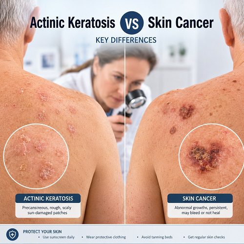
When comparing actinic keratosis vs skin cancer, one of the most important points to remember is that actinic keratosis is usually considered a precancerous skin lesion, whereas skin cancer involves abnormal cells that have become cancerous. However, not every actinic keratosis will become cancer.
Both conditions can appear as changes on sun-exposed areas such as the face, scalp, ears, neck, forearms and hands. Some lesions may look similar in their early stages, making it difficult for someone without medical training to determine what a particular skin change represents.
The key differences between actinic keratosis and skin cancer, including their appearance, symptoms, causes, risk factors, diagnosis, treatment and warning signs that should prompt medical attention.
What is Actinic Keratosis?
What Is Actinic Keratosis? Actinic keratosis is a common skin condition caused primarily by cumulative exposure to ultraviolet radiation from sunlight or tanning beds. It appears as a rough, scaly or crusty patch on areas of skin that receive significant sun exposure.
Actinic keratosis is often described as a precancerous skin lesion because some lesions can progress to squamous cell carcinoma, a type of skin cancer. However, progression does not occur in every case.
Lesions can vary in colour and appearance. They may be pink, red, brown, tan or close to the person’s natural skin tone. The surface often feels rough or sandpaper-like, sometimes before the lesion becomes clearly visible.
Common locations include:
- Face and forehead
- Ears
- Scalp, particularly in people with little or no hair
- Neck
- Forearms
- Back of the hands
- Shoulders
- Lower legs in some people
Actinic keratoses are particularly common in older adults and people who have accumulated substantial sun exposure throughout their lives.
What Is Skin Cancer?
Skin cancer occurs when abnormal skin cells grow uncontrollably. There are several types of skin cancer, with the most common being basal cell carcinoma and squamous cell carcinoma. Melanoma is less common but can be more aggressive.
Skin cancer can develop on sun-exposed skin, although it may also occur in areas that receive little sunlight.
The appearance of skin cancer varies depending on the type. It may present as a new growth, a sore that does not heal, a changing mole, a scaly patch, a pearly bump or a lesion that repeatedly bleeds or develops a crust.
Because skin cancer can sometimes resemble harmless skin conditions, persistent or changing lesions should be assessed by a healthcare professional.
Actinic Keratosis vs Skin Cancer: Key Differences
The main difference between these conditions is the nature of the abnormal cells.
Actinic keratosis involves damaged skin cells that have developed because of cumulative UV exposure. Some lesions can progress to cancer, particularly squamous cell carcinoma.
Skin cancer, on the other hand, occurs when abnormal cells have acquired changes that allow them to grow and behave as cancerous cells.
Here are some important differences:
| Feature | Actinic Keratosis | Skin Cancer |
| Nature | Precancerous or potentially precancerous lesion | Cancerous growth |
| Main cause | Long-term UV exposure | Multiple factors, including UV exposure and genetic changes |
| Appearance | Usually rough, scaly patch | May be a growth, ulcer, changing mole or persistent lesion |
| Symptoms | May be rough, itchy, tender or asymptomatic | May bleed, ulcerate, hurt, itch or change |
| Progression | Some lesions may become squamous cell carcinoma | Can grow and potentially spread depending on type |
| Diagnosis | Clinical examination and sometimes biopsy | Examination and biopsy are commonly used |
| Treatment | Freezing, topical medicines, photodynamic therapy and other methods | Surgery, topical treatment, radiation, immunotherapy or other treatments depending on type |
Although these differences can be helpful, they cannot replace professional assessment. A lesion that looks like actinic keratosis may occasionally represent skin cancer.
How Does Actinic Keratosis Look?
Recognising the typical appearance of actinic keratosis can help you identify skin changes that deserve attention.
An actinic keratosis may appear as:
- A rough or scaly patch
- A dry, crusty area
- A flat or slightly raised lesion
- A pink, red, brown or skin-coloured patch
- A lesion that feels rougher than the surrounding skin
- A small area that repeatedly flakes or develops a crust
Some actinic keratoses are easier to feel than to see. Running a fingertip gently over the skin may reveal a rough patch even when the colour difference is subtle.
Lesions can be very small, but they may gradually become larger or thicker.
What Are Common Skin Cancer Signs?
Recognising skin cancer signs is important because early diagnosis can improve treatment options and outcomes.
The appearance depends on the type of cancer.
Basal Cell Carcinoma
Basal cell carcinoma may appear as:
- A pearly or shiny bump
- A pink or flesh-coloured growth
- A sore that repeatedly heals and returns
- A lesion that bleeds easily
- A flat, scaly or slightly depressed patch
Squamous Cell Carcinoma
Squamous cell carcinoma may appear as:
- A rough, scaly patch
- A firm raised growth
- A crusted lesion
- A sore that does not heal
- A lesion that bleeds or becomes tender
Because squamous cell carcinoma can sometimes develop from actinic keratosis, a lesion that becomes thicker, more painful, ulcerated or persistently inflamed should be assessed.
Melanoma
Melanoma commonly develops from pigment-producing cells. It can appear as a new dark spot or a mole that changes.
The ABCDE approach can help identify suspicious pigmented lesions:
A – Asymmetry: One half does not match the other.
B – Border: The edges are irregular, uneven or poorly defined.
C – Colour: The lesion contains multiple or unusual colours.
D – Diameter: The lesion is larger than expected, although melanoma can be smaller.
E – Evolving: The mole or spot changes in size, shape, colour or symptoms.
Not every melanoma follows the ABCDE pattern, so any concerning or changing lesion should be evaluated.
Can Actinic Keratosis Turn Into Skin Cancer?
Yes, some actinic keratoses can progress to squamous cell carcinoma. However, the risk for an individual lesion varies, and many actinic keratoses never become cancerous.
Actinic keratosis is therefore best viewed as a warning sign of significant sun damage rather than an indication that someone already has skin cancer.
Having multiple actinic keratoses can indicate substantial cumulative UV damage. People with these lesions may also have an increased risk of developing other forms of skin cancer.
For this reason, monitoring the skin and attending recommended dermatology appointments can be important.
Why Does UV Exposure Matter?
UV radiation is one of the most important environmental risk factors for actinic keratosis and many forms of skin cancer.
Repeated exposure can damage DNA within skin cells. Over many years, this damage can accumulate and contribute to abnormal cell growth.
Risk can increase with:
- Frequent sun exposure
- History of sunburn
- Outdoor occupations or hobbies
- Use of tanning beds
- Fair skin
- Older age
- Previous actinic keratoses
- Previous skin cancer
- A weakened immune system
UV exposure is not limited to particularly hot or sunny days. Ultraviolet radiation can still affect the skin when the weather is cooler or partly cloudy.
Who Is More Likely to Develop Actinic Keratosis?
Actinic keratosis is particularly common among people with long-term sun exposure. It becomes more frequent with age because UV-related damage accumulates over time.
People at higher risk may include those who:
- Spend considerable time outdoors
- Have fair or sun-sensitive skin
- Have had repeated sunburns
- Live in areas with strong UV exposure
- Have used tanning beds
- Have a history of actinic keratosis
- Have previously developed certain types of skin cancer
- Take medicines or have conditions that weaken immune function
However, people with darker skin can also develop actinic keratosis and skin cancer. Everyone should pay attention to persistent or changing skin lesions.
How Are Actinic Keratosis and Skin Cancer Diagnosed?
A healthcare professional usually begins with a visual and physical examination of the skin.
For suspected actinic keratosis, a dermatologist may be able to make a diagnosis based on the lesion’s appearance and texture. A dermatoscope may also help the clinician examine features that are not visible to the naked eye.
If the lesion looks unusual, thick, persistent or suspicious for cancer, a biopsy may be recommended. During a biopsy, a small sample of tissue is removed and examined under a microscope.
A biopsy can help determine whether a lesion represents actinic keratosis, squamous cell carcinoma, basal cell carcinoma, melanoma or another skin condition.
Treatment for Actinic Keratosis
Actinic Keratosis Treatment depends on the number of lesions, their location, thickness and other individual factors.
Common approaches include:
Cryotherapy
Cryotherapy uses extreme cold, usually liquid nitrogen, to destroy the abnormal cells. The treated area may blister or crust before healing.
Prescription Topical Treatments
Certain prescription creams or gels can be applied to areas affected by actinic keratoses. They work by targeting abnormal or damaged cells.
Photodynamic Therapy
Photodynamic therapy uses a light-sensitive medication followed by a specific light treatment to destroy abnormal cells.
Curettage or Other Procedures
For thicker or particularly suspicious lesions, a clinician may remove the abnormal tissue using a procedure such as curettage or another surgical technique.
The best treatment depends on the individual and should be determined by a qualified healthcare professional.
How Is Skin Cancer Treated?
Skin cancer treatment depends heavily on the type, size, location and stage of the cancer.
For many early skin cancers, surgical removal is the primary treatment. Procedures may include standard excision or Mohs surgery for selected cancers.
Other treatments can include:
- Topical medicines for certain superficial cancers
- Curettage and electrosurgery
- Radiation therapy
- Immunotherapy
- Targeted therapies
- Other systemic treatments for advanced disease
Melanoma treatment can differ significantly from treatment for basal or squamous cell carcinoma, particularly when melanoma has spread.
Early diagnosis generally makes treatment more straightforward, which is why suspicious lesions should not be ignored.
When Should You See a Doctor?
It is sensible to arrange a medical assessment if you notice a skin lesion that:
- Does not heal
- Continues to grow
- Becomes thicker
- Bleeds repeatedly
- Develops an ulcer
- Becomes painful or unusually tender
- Changes in colour or shape
- Develops an irregular border
- Looks significantly different from surrounding lesions
- Persists despite basic skin care
A lesion that was previously diagnosed as actinic keratosis should also be reassessed if it changes significantly.
Do not attempt to diagnose skin cancer solely from photographs or online descriptions. Several benign, precancerous and cancerous skin conditions can look alike.
Can Actinic Keratosis Be Prevented?
You cannot reverse all previous UV damage, but reducing further exposure can help protect your skin.
Practical sun-protection measures include:
- Seek shade when UV levels are high.
- Wear protective clothing when spending extended time outdoors.
- Use a broad-spectrum sunscreen as directed.
- Reapply sunscreen as recommended, particularly after swimming or sweating.
- Wear a wide-brimmed hat and UV-protective sunglasses.
- Avoid tanning beds.
- Examine your skin regularly for new or changing lesions.
Sun protection should be part of a long-term routine rather than something used only during holidays.
Actinic Keratosis vs Skin Cancer: Why Early Assessment Matters
The most important lesson when considering actinic keratosis vs skin cancer is that appearance alone is not always enough to distinguish them.
Actinic keratosis represents UV-related damage and may be considered a precancerous skin lesion. Skin cancer involves malignant cell growth. Some skin cancers can resemble actinic keratosis, particularly squamous cell carcinoma.
Therefore, a persistent rough patch should not automatically be assumed to be harmless. Similarly, being diagnosed with actinic keratosis does not mean that cancer is inevitable.
Regular skin checks can help identify changes early. If a lesion changes, becomes thicker, starts bleeding or develops other unusual characteristics, a dermatologist or other qualified healthcare professional can determine whether further investigation is needed.
Frequently Asked Questions
Is actinic keratosis skin cancer?
No. Actinic keratosis is generally considered a precancerous or potentially precancerous lesion rather than skin cancer. Some lesions can progress to squamous cell carcinoma.
What is the main difference between actinic keratosis and skin cancer?
Actinic keratosis involves abnormal, sun-damaged skin cells, while skin cancer involves malignant cell growth. However, some early skin cancers can look similar to actinic keratosis.
Is actinic keratosis dangerous?
Actinic keratosis is not usually an immediate medical emergency, but it should not be ignored. Some lesions can develop into squamous cell carcinoma, and people with multiple actinic keratoses may have significant cumulative sun damage.
Can actinic keratosis go away on its own?
Some lesions may become less noticeable, but this does not necessarily mean the underlying sun damage has disappeared. A healthcare professional can advise whether monitoring or treatment is appropriate.
Should every actinic keratosis be treated?
Treatment decisions depend on the lesion, the person’s circumstances and the clinician’s assessment. A dermatologist can recommend whether treatment, monitoring or further investigation is appropriate.
Final Thoughts
Understanding actinic keratosis vs skin cancer can help you recognise why persistent skin changes deserve attention. Actinic keratosis is commonly associated with long-term UV damage and is considered a precancerous skin lesion, while skin cancer develops when abnormal cells become malignant.
The two conditions can sometimes look alike, particularly during their early stages. Rough, scaly patches, non-healing sores, bleeding lesions, changing moles and other persistent skin abnormalities should therefore be assessed rather than self-diagnosed.
Protecting your skin from excessive UV exposure, checking for new or changing lesions and seeking professional advice when something looks unusual are important parts of skin health.
Medical disclaimer
This article is intended for general educational purposes and does not replace professional medical advice, diagnosis or treatment. If you have a new, changing, bleeding, painful or persistent skin lesion, consult a qualified healthcare professional or dermatologist for an appropriate assessment.